1602
A Novel Asymmetric 16-Element pTx Transceiver Coil Array: Towards Denser Elements for Improved RF-Shimming and G-Factor for Parallel Cardiac MRI in Pigs at 7T1Chair of Cellular and Molecular Imaging, Comprehensive Heart Failure Center (CHFC), University Hospital Wuerzburg, Wuerzburg, Germany
Synopsis
A novel 8Tx/16Rx coil array was designed, simulated, and tested in phantom and for cardiac magnetic resonance imaging (cMRI) ex-vivo pigs at 7T. The 16-elements of the array were distributed on a half-elliptical shape housing. Combined signal-to-noise ratio (SNR) maps and FA-maps were acquired using the developed coil before and after RF-shimming. The novel cardiac array supports parallel imaging with acceleration factors of up to R=4 without a significant degradation in the image quality. High-resolution ex-vivo cardiac images were acquired with 0.3mm x 0.3mm in plane resolution. The dedicated coil enhances the SNR within the heart by about six-times compared to a commercial human cardiac coil array.
Introduction
Increasing the number of elements in the coil array improves the SNR, the geometry factors (g-factor) and the B1+-field homogeneity for parallel imaging. However, the coupling between the resonant elements will increase and peak splitting will occur and this issue will limit the possibility to control the shape of the individual B1+-fields generated from each element within the heart. The decoupling mechanism and the distribution of elements on the array is an important aspect to minimize coupling and hence for efficient RF-shimming 1-3 and parallel imaging with high g-factors. Performing cMRI in swine plays an important role in the development of new MRI methodology at 7T. However, pigs have quite different thorax shape than humans. The misuse of the human coils for performing cMRI in pigs will lead to a degradation of the SNR and B1+-field homogeneity due to less loading and suboptimal coil dimensions. Different types of coil arrays have been developed for body imaging at UHF 7T and 10.5T, such as local multichannel loops 4-7 and dipole antenna arrays 8-10. Building a dedicated cardiac coil array for pigs could be considered as an important step toward having an optimized multichannel pTx coil array for humans at 7T.Materials and Methods
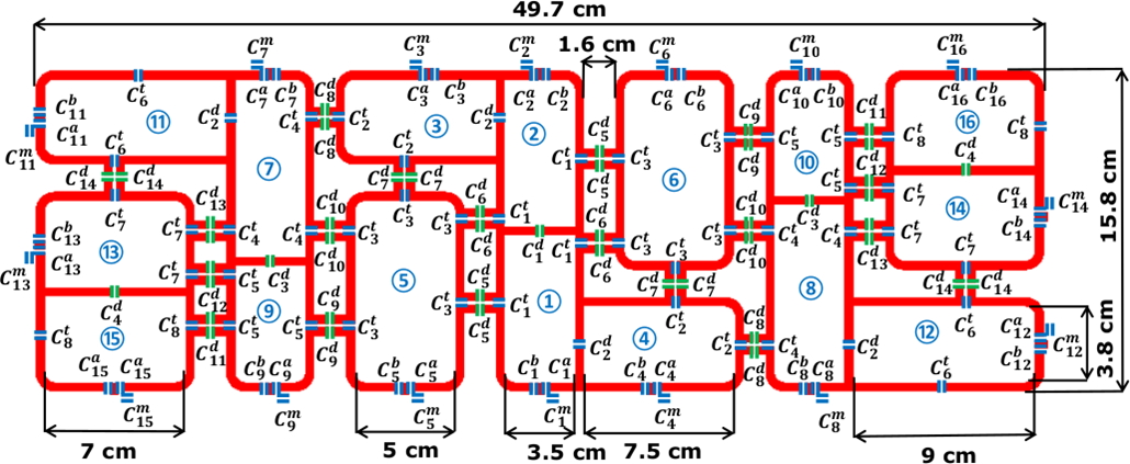
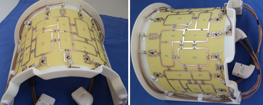
The novel design is composed of 16-elements and they were arranged in such a way that each seven-elements are reversed and mirrored around the central two-elements. The size of the central two-elements 1 and 2 was 3.5×7.3 cm2. The decoupling between the central two-elements was accomplished using a common conductor and a shared decoupling capacitor (C1d) 11.The other 14-elements were distributed around the central two-loops in a reversed and mirrored way to allow good decoupling between all loops and a varying B1+-field distribution (Figure 1). The size of the elements 3 and 4 was 3.8×7.5 cm2. The elements 3 and 4 were reversed and decoupled with elements 1 and 2 using a shared decoupling capacitor (C2d). The size of the elements 5 and 6 was 5×9 cm2. The elements 7 and 9 and the reversed identical elements 8 and 10 were decoupled using a shared decoupling capacitor (C3d). The elements 1, 2, 3, and 4 were decoupled from the neighboring elements 5 and 6 using capacitive decoupling (C5d, C6d and C7d) in addition to a decoupling gap of 1.6 cm and 1.1 cm. The total external dimension for the array was 15.8×49.7 cm2 ( Figure 1). EM-simulations were performed using CST-Microwave-Studio for coil design and B1+-field calculations. The copper track width is 4 mm etched on a 0.3 mm FR4-PCB. All feeding ports, tuning, matching, and decoupling capacitors were modeled as 50-Ω discrete face ports 12. The final total number of mesh cells is 38.3 million. All measurements were performed on a 7T whole-body MAGNETOM Siemens Terra scanner equipped with a 16-kW RF amplifier for 8-channel pTx mode. The novel coil was designed to have one half-elliptical shape housing (Xrad/Yrad=17/21 cm) to demonstrate the feasibility of performing cMRI for pigs (Figure 2). The novel coil design was tested in phantom and 46-kg ex-vivo pig cMRI with the measurement of SNR maps, B1-shimming, g-factor with high parallel imaging acceleration factors.Results and Discussion
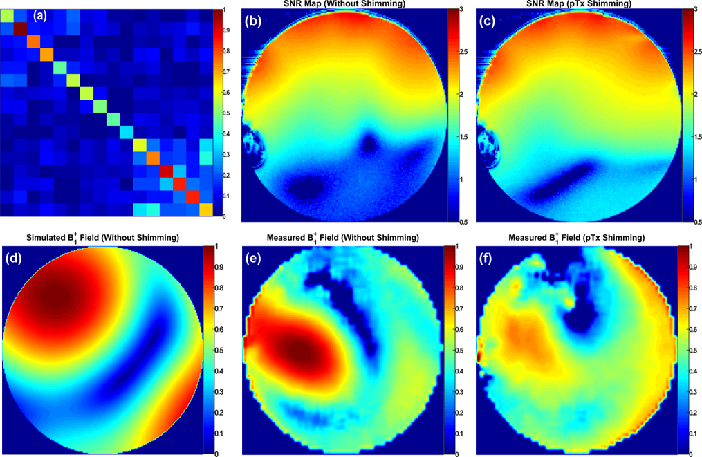
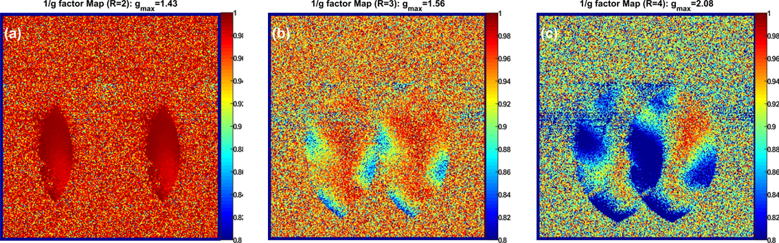
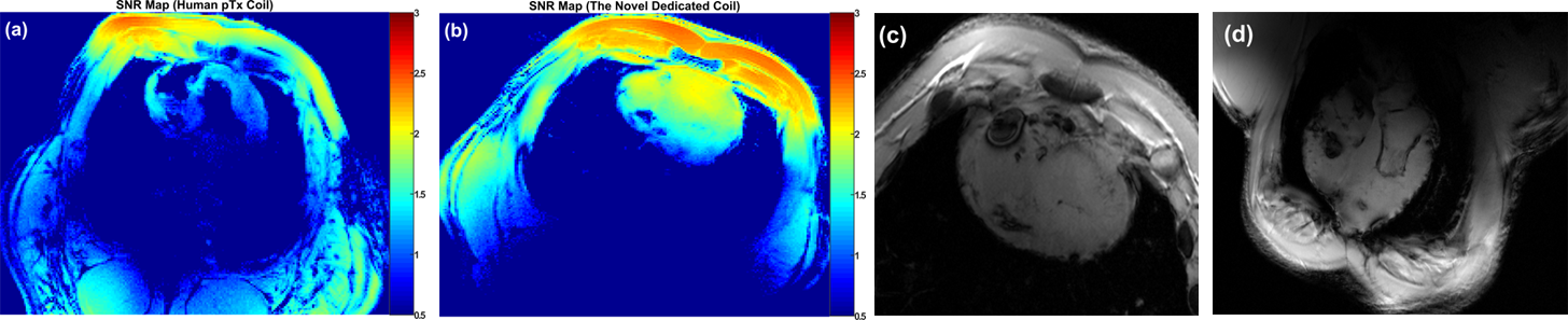
Figure 3 demonstrates the results of the phantom MR-measurements, the noise covariance matrix, the SNR-maps performed before and after RF-shimming. The coil shows a mean of the SNR of 117 compared to 112 before RF-shimming. With the implemented phases and even prior RF-shimming, the coil shows good Rx-properties with high SNR. The coil shows a significant improvement in the B1+-field homogeneity after RF-shimming (Figure 3f) which proves the high efficiency of the developed array design for pTx-systems. For fair comparison, the combined B1+-field was normalized to its own maximum. The simulated and measured coronal B1+-field distribution (Figure 3d and e) shows some agreements in the local maxima and destructive interference locations. For better agreement, the amplitudes of all elements need to be calibrated based on the S-matrix and FA-map measurements. Parallel imaging performance were measured using acceleration factors R=2, 3, and 4 (Figure 4). The mean SNR within the heart region from the novel design was 61.6 compared to 10 from a commercial human coil (Figure 5b). The developed coil demonstrates essentially better B1+-field homogeneity within heart without a significant destructive interference (Figure 5). High-resolution ex-vivo images with in-plane pixel resolution of 0.3 mm x 0.3 mm were acquired from the dedicated pig coil.Conclusion
The novel asymmetric 8Tx/16Rx coil array has demonstrated good decoupling provided by reversed geometrical loop orientations and improved control of the B1+-field distributions with more degrees of freedom for RF-shimming. The developed 8Tx/16Rx cardiac array demonstrated high efficiency in both Tx and Rx properties for cMRI at 7T.Acknowledgements
Financial support: German Ministry of Education and Research (BMBF, grants: 01EO1004, 01E1O1504).
The animals were provided for ex-vivo measurements after they were euthanized following their approved use in project 55.2 DMS 2532-2-664 (Regierung Unterfranken). Euthanasia was performed with intravenous application of 150 mg/kg pentobarbital under isoflurane anesthesia with fentanyl analgesia.
References
- Xin, S. X., Huang, Q., Gao, Y., et al., (2013). Fetus MRI at 7 T: Shimming Strategy and SAR safety implications. IEEE Transactions on Microwave Theory and Techniques, 61, 2146-2152.
- Ibrahim, T. S. (2006). Ultrahigh-field MRI whole-slice and localized RF field excitations using the same RF transmit array. IEEE transactions on medical imaging, 25(10), 1341-1347.
- Mao, W., Smith, M. B., and Collins, C. M. (2006). Exploring the limits of RF shimming for high-field MRI of the human head. Magnetic Resonance in Medicine, 56(4), 918-922.
- Graessl, A., Renz, W., Hezel, F., et al., (2014). Modular 32-channel transceiver coil array for cardiac MRI at 7.0 T. Magnetic resonance in medicine, 72(1), 276-290.
- Dieringer, M. A., Renz, W., Lindel, T., et al., (2011). Design and application of a four-channel transmit/receive surface coil for functional cardiac imaging at 7T. Journal of Magnetic Resonance Imaging, 33(3), 736-741.
- Gräßl, A., Winter, L., Thalhammer, C., et al., (2013). Design, evaluation and application of an eight channel transmit/receive coil array for cardiac MRI at 7.0 T. European journal of radiology, 82(5), 752-759.
- Thalhammer, C., Renz, W., Winter, L., et al., (2012). Two-dimensional sixteen channel transmit/receive coil array for cardiac MRI at 7.0 T: design, evaluation, and application. Journal of Magnetic Resonance Imaging, 36(4), 847-857.
- Oezerdem, C., Winter, L., Graessl, A., et al., (2016). 16-channel bow tie antenna transceiver array for cardiac MR at 7.0 tesla. Magnetic resonance in medicine, 75(6), 2553-2565.
- Raaijmakers, A. J. E., Luijten, P. R., and van Den Berg, C. A. (2016). Dipole antennas for ultrahigh-field body imaging: a comparison with loop coils. NMR in Biomedicine, 29(9), 1122-1130.
- Ertürk, M. A., Wu, X., Eryaman, Y., Van de Moortele, P. F., et al., (2017). Toward imaging the body at 10.5 tesla. Magnetic resonance in medicine, 77(1), 434-443.
- Perrier, A. L., Grenier, D., Pouzin, A., et al., (2012). Design of a two-channel NMR coil using an impedance transformation approach. IEEE Sensors Journal, 12(6), 1801-1808.
- Kozlov, M., and Turner, R. (2009). Fast MRI coil analysis based on 3-D electromagnetic and RF circuit co-simulation. Journal of magnetic resonance, 200(1), 147-152.
Figures