1597
Rapid Material Characterization For 3D-printed MRI Coils: A Deep Learning Approach1Electrical Engineering, University of Wisconsin-Madison, Madison, WI, United States, 2Radiology, University of Wisconsin-Madison, Madison, WI, United States, 3Medical Physics, University of Wisconsin-Madison, Madison, WI, United States
Synopsis
A novel material characterization approach which is based on a microstrip line test fixture paired with deep learning analysis, is presented to optimize the use of additive manufacturing (3D-printing) in constructing the structure of MRI coils with arbitrary 3D geometries. This type of manufacturing is appealing because it can be used to construct geometries that are time-consuming and expensive to make using traditional machining methods. Full-wave electromagnetic simulations are efficient and promising technique to expedite the design process of MRI coils and therefore, it is crucial to include the electrical properties of 3D-printed materials in the electromagnetic simulations because it affects coil performance.
Purpose
3D-printing is a low-cost and rapid means to construct MRI coils with arbitrary 3D geometries [1]–[3]. RF coil design in MRI requires the knowledge of electromagnetic field distribution. This is achieved now by using full-wave electromagnetic (EM) simulations. To simulate an optimized coil, it is necessary to include the material properties (e.g., dielectric constant, loss tangent) of the coil components because the response of a non-magnetic material to EM waves depends primarily on the dielectric constant and loss tangent of the material. 3D-printed polymers are non-standardized materials and their electrical properties are likely to vary between manufacturers, which can affect coil performance. There are many approaches to measure dielectric properties of materials [4]. However, all of these approaches have limitations for characterizing 3D-printed polymers at MRI relevant frequencies. In recent work [3], a test fixture for material characterization was proposed utilizing EM simulations along with the S-parameter measurements to characterize the dielectric properties of 3D-printed materials at MRI related frequencies. This method of characterization, although achieving accurate results, can be very time-consuming due to the computational requirements of EM simulations. Using deep learning approaches can reduce the computational time significantly. The goal of this study is to propose a broadband (applicable to MRI frequencies) and simple to build test fixture applicable to 3D-printed materials and use the power of deep learning to perform rapid material characterization and find accurate results from the estimated scattering parameters.Methods
The proposed test fixture is a microstrip transmission line terminated with a parallel plate capacitor. The material under test (MUT) serves as the dielectric between the capacitor plates as shown in Figure 1. Any change in the dielectric constant or loss inside the MUT results in a change in the magnitude and phase of the reflection coefficient (S11) measured by a vector network analyzer (VNA) connected to the microstrip transmission line. This change was used to characterize the material between the capacitor plates. EM simulations of the test fixture were developed and performed in CST Microwave Studio using frequency and parameter sweep solvers with two parameters: MUT dielectric constant and MUT loss tangent. The range of simulated dielectric constant (relative permittivity) was from 1 to 5 which is an expected range for polymers and from 0.001 to 0.05 for loss tangent. Because 3D-printed materials are typically low-dispersive, the changes in their dielectric properties are negligible in the simulated range of frequencies and can be assumed independent of frequency [4]. The dataset consisted of 2091 S11 calculated from EM simulations, each with 200 frequency samples in the range of 5-500 MHz. The data was processed in Python using Tensorflow. Data was split into three sets: training (1504 data), validation (377 data), and testing (210). Figure 2 shows the neural network used in this study. The Adam optimizer and mean squared error (MSE) loss were used to train the model. To achieve better performance in predicting the loss tangent values the input data was represented with magnitude in dB scale, real, and imaginary values because the small changes in magnitude of S11 due to a change in loss tangent was much more distinguishable in the magnitude in dB scale.Results
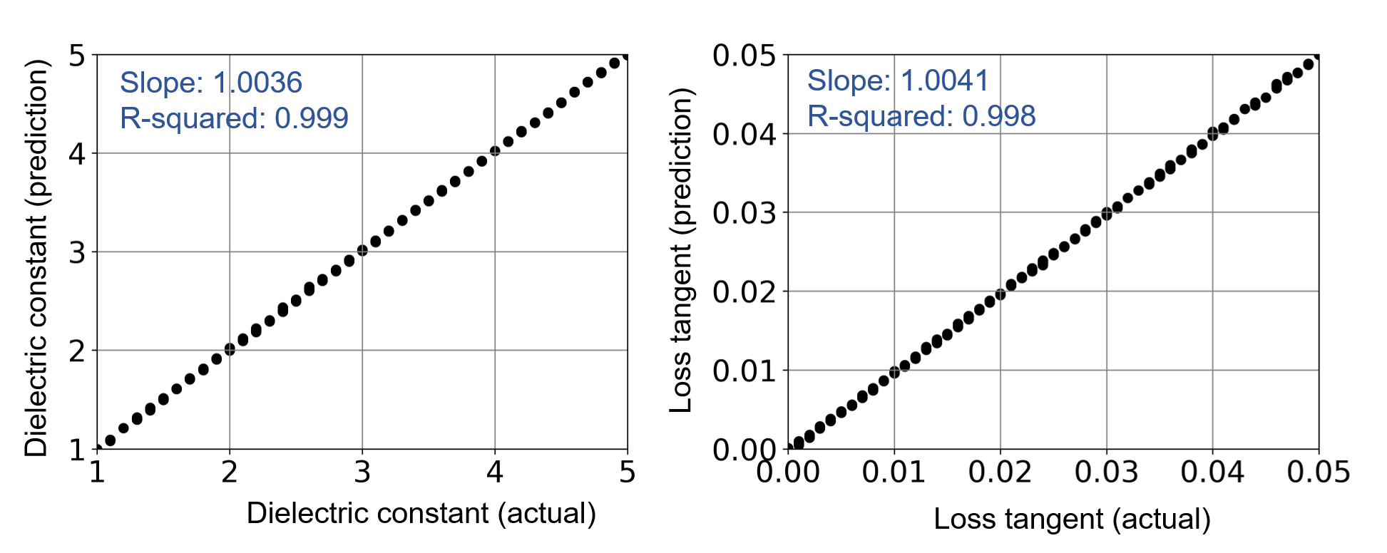
Figure 3 shows different coils constructed based on 3D-printing. Figure 4 shows the predicted values vs. actual values for dielectric constant and loss tangent. The r-squared value for prediction of dielectric constant was 0.999 and was 0.998 for loss tangent which demonstrates strong performance of our model.Discussion and Conclusion
Additive manufacturing has provided new means to accurately construct patient/anatomy-specific medical devices. The use of this type of manufacturing in the development of MRI devices is highly appealing and It is important to measure electrical properties of the 3D-printed materials before using in MRI RF coils because of the potential effects they might have on the coil performance. The actual 3D field profile of a coil depends on the response of all materials used in the coil and it is a less than ideal time for modifications to be made after the coil was built. This abstract, demonstrates a rapid approach applicable to 3D-printed materials and MRI frequencies to measure the dielectric properties of dielectric materials by utilizing deep learning to perform estimation of electrical properties of materials from an easy to build test fixture (which cannot be readily analyzed using conventional approaches).Acknowledgements
No acknowledgement found.References
[1] S. Wei, Z. Wang, H. Wang, X. Lyu, L. Deng, and W. Yang, “Design and implementation of MRI RF coil based on 3D printing,” in 2015 IEEE MTT-S 2015 International Microwave Workshop Series on RF and Wireless Technologies for Biomedical and Healthcare Applications (IMWS-BIO), 2015, pp. 222–224.
[2] K.-H. Herrmann, C. Gärtner, D. Güllmar, M. Krämer, and J. R. Reichenbach, “3D printing of MRI compatible components: Why every MRI research group should have a low-budget 3D printer,” Med. Eng. Phys., vol. 36, no. 10, pp. 1373–1380, Oct. 2014.
[3] B. Behzadnezhad, B. D. Collick, N. Behdad, and A. B. McMillan, “Dielectric properties of 3D-printed materials for anatomy specific 3D-printed MRI coils,” J. Magn. Reson., vol. 289, pp. 113–121, Apr. 2018
[4] P. I. Deffenbaugh, R. C. Rumpf, and K. H. Church, “Broadband Microwave Frequency Characterization of 3-D Printed Materials,” IEEE Trans. Components, Packag. Manuf. Technol., vol. 3, no. 12, pp. 2147–2155, Dec. 2013.
Figures