1596
Solid material resembling human tissues:a white and gray matter brain phantom.1Department of Electrical Engineering, Pontificia Universidad Católica de Chile, Santiago, Chile, 2Biomedical Imaging Center, Pontificia Universidad Católica de Chile, Santiago, Chile, 3Millennium Nucleus for Cardiovascular Magnetic Resonance, Santiago, Chile, 4Faculty of Chemistry, Deparment of Organic Chemistry, Pontificia Universidad Católica de Chile, Santiago, Chile, 5Department of Radiology, Pontificia Universidad Católica de Chile, Santiago, Chile
Synopsis
In this work we present a solid brain phantom that resemble the anatomy and T2 relaxation times of the brain. The developed material is a 3-component mix. Adjusting the relative concentration of the compounds allow to modulate T2 relaxation time, following a linear relationship within a range of 145 to 263ms. The resulting solid phantom can reproduce correctly the geometry of white and gray matter. The range of achievable T2 relaxation times makes possible the construction of phantoms that could mimic a wide range of biological tissues.
Introduction
Physical phantoms are commonly used for quality assurance (QA) and calibrating purposes. Existing phantoms (e.g. ADNI Magphan®1, 2, or NIST Phannie3, 4) can mimic relaxation times, but they have simple geometrical shapes, Other phantoms can simulate more realistic anatomies5-8, but they are made of only one contrast media, thus they cannot represent heterogeneous structures. In this work, we present a solid MRI phantom capable of resembling the shape and T2 relaxation times of a two-compartment brain (gray/white matter). The phantom is made of mixtures of 3 compounds: dimethyl siloxane, as the base component; silicone thinner, that lower the viscosity of the mix thus facilitating to pour it into a mold and to remove air bubbles from it; and a polyether named “Q” that allow modulating T2 relaxation time.Methodology
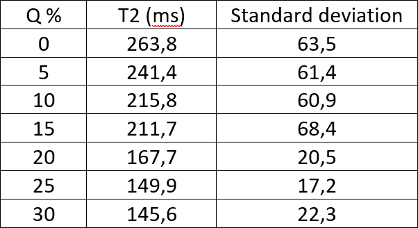
Mixtures were produced using the base material and thinner (ratio 90:10). Q was added to the base within a range of 0-30% of the total weight. Empirical tests showed us that the amount of Q is inversely proportional to T2. Above 30%, the Q mixture becomes viscous and making harder to remove air bubbles and to pour it into the molds. To analyze the Q-concentration vs. T2 relationship, we built 7 cylinders with Q concentration [0, 5, 10, 15, 20, 25, 30]%. T2 values of the samples were characterized using a spin echo sequence with 8 echoes (TR=2000, TE every 25ms, voxel size 0.28x0.28x4mm) using a Philips 1.5T Achieva scanner and in-house Matlab-based software.
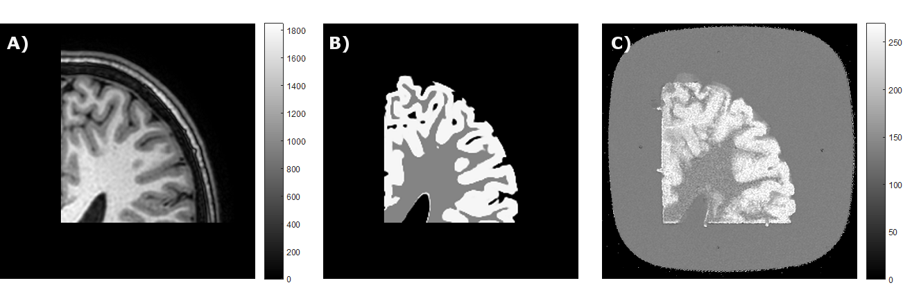
Two mixtures were used to create a brain phantom with two tissues.
Gray/white matter were segmented using SPM129 from
a T1 weighted image of a volunteer. As a proof of concept, we considered only a
portion of the left parietal lobe. Gray/white matter molds were created using a
3D printer (Prusa I3) with 0,2mm layers. White matter was made of a 0%
Q-mixture, so that to match approximately a T2 of 260ms. The mixture was poured into a mold inside a
vacuum chamber to remove air bubbles and it was left curing for a day. White
matter was removed and placed into a second mold. Grey matter was made of a 20%
Q-mixture, so that to match a approximately T2 of 170ms. Pouring this second mixture
followed the same procedure previously described. T2 characterization of the
phantom followed the same previous procedure.
Results
T2 followed a quasi-linear relationship with Q concentration (Table1), following a range between 145.6 and 263.8ms. The solid brain phantom resembles correctly the geometry of gray and white matter with the desired T2 relaxation times (Fig1).
Discussion
Having a solid phantom opens a great opportunity for QA, scanner calibration and sequence development. Existing liquid or gel-based phantoms have some problems in terms of stability, motion artifacts, and the restriction to create different tissues. The modulated T2 range is reasonably large to resemble a wide variety of biological tissues. Our current material can modulate T1 in the range [600-700]ms, we are exploring some alternatives to widen this range by adding additional compounds to the mixtures. The proliferation of air bubbles is probably the main restriction for this T1 modifier search.Conclusion
We developed a material and a methodology suitable for building solid phantoms that can resemble correctly the geometry and T2 times of complex structures such as a brain. This solid material can mimic a wide range of T2 times, thus may be used to create phantoms of a large variety of biological structures.Acknowledgements
This publication has received funding from Conicyt Fondecyt 1161448, Millenium Science Initiative of the Ministry of Economy, Development and Tourism, grant Nucleus for Cardiovascular Magnetic Resonance, CONICYT, PIA-ACT1416), and CONICYT-PCHA/Doctorado Nacional/2015 – 21151003
References
1. Qiu, J., et al., Design of multi-function MR test phantom. Chin. J. Med. Imaging Technol, 2012. 28: p. 997-1000.
2. Li, J.-Y., et al., Comparison of MR Imaging Quality Parameters between Spin Echo and Fast Spin Echo Imaging Pulse Sequence. Chinese Medical Equipment Journal, 2012. 4: p. 058.
3. Selwyn, R., Phantoms for Magnetic Resonance Imaging, in The Phantoms of Medical and Health Physics: Devices for Research and Development, A.L. DeWerd and M. Kissick, Editors. 2014, Springer New York: New York, NY. p. 181-199.
4. Russek, S.E., et al. Characterization of NIST/ISMRM MRI system phantom. in Proc. Intl. Soc. Mag. Reson. Med. 2012.
5. Chen, S.J.-S., et al., An anthropomorphic polyvinyl alcohol brain phantom based on Colin27 for use in multimodal imaging. Medical Physics, 2012. 39(1): p. 554-561.
6. Saotome, K., et al., A brain phantom for motion-corrected PROPELLER showing image contrast and construction similar to those of in vivo MRI. Magnetic Resonance Imaging, 2017. 36: p. 32-39.
7. Surry, K.J.M., et al., Poly(vinyl alcohol) cryogel phantoms for use in ultrasound and MR imaging. Physics in Medicine and Biology, 2004. 49(24): p. 5529.
8. Kim, K.S., et al., A Compressed-Sensing Based Blind Deconvolution Method for Image Deblurring in Dental Cone-Beam Computed Tomography. Journal of Digital Imaging, 2018.
9. in Statistical Parametric Mapping, K. Friston, et al., Editors. 2007, Academic Press: London.
Figures