1595
A 3T MRI platform for imaging rodent models by integrating a dedicated high-strength gradient coil on a whole-body magnet1Institute of Biomedical Engineering and Nanomedicine, National Health Research Institutes, Miaoli, Taiwan, 2Department of Biomedical Engineering and Environmental Sciences, National Tsing Hua University, Hsinchu, Taiwan, 3Institute of Neuroscience and Medicine 4, INM-4, Forschungszentrum Jülich, Jülich, Germany, 4Institute of Neuroscience and Medicine 11, INM-11, JARA, Forschungszentrum Jülich, Jülich, Germany, 5JARA - BRAIN - Translational Medicine, Aachen, Germany, 6Department of Neurology, RWTH Aachen University, Aachen, Germany, 7MR Solutions Ltd., Guildford, United Kingdom, 8Department of Medical Research, Chi Mei Medical Center, Tainan, Taiwan, 9Institute of Medical Device and Imaging, National Taiwan University College of Medicine, Taipei, Taiwan
Synopsis
In this study, a high-strength gradient coil dedicated for imaging rodent models has been successfully integrated on a whole-body 3T MRI magnet. The imaging capability of this system has been qualitatively and quantitatively demonstrated using phantom, ex-vivo specimen and in-vivo rat experiments. Since the hardware components used for integration on our system are independent from system vendors, this study can be a practically useful guide, especially for those who want to conduct small animal imaging on clinically used magnets.
Introduction
Although studying animal models on ultra-high field MRI is beneficial to understand the underlying mechanisms of disease, the translational use of developed sequences or protocols is sometimes difficult due to the incompatible field strengths on most of the clinical MRI scanners. Therefore, an alternative and efficient approach is to develop an inserted gradient coil system on clinical MRI magnet and perform the compatible imaging sequences on the same magnet. Previous literature has shown some strategies to conduct the small animal imaging on clinical MRI systems, with either original equipped coils or inserted gradient coil and customized radio-frequency (RF) coils 1-7. In this study, we aimed to integrate a dedicated high-strength gradient coil on a whole-body 3T magnet. The performance was assessed qualitatively and quantitatively by using dedicated phantoms, and the capability to perform advanced imaging sequences, e.g. diffusion tensor imaging (DTI) and quantitative susceptibility mapping (QSM), was examined on ex-vivo specimen as well as in-vivo rat models.Methods
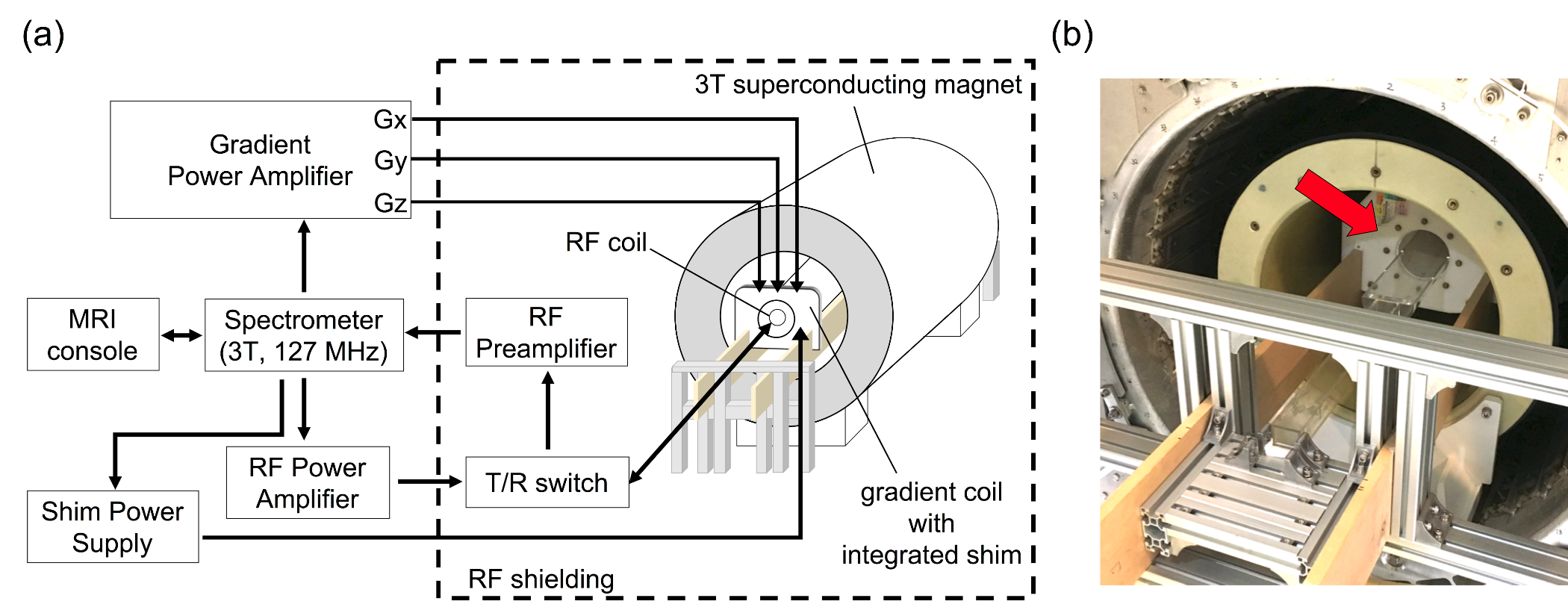
A whole-body 3T magnet was adapted as the main platform on our system. A high-strength gradient coil with maximum strength of 675 mT/m and integrated shim channels (Resonance Research Inc., USA) was installed and mechanically fixed by using wooden- and aluminum-made structures.The gradient coil was driven by a commercially available gradient power amplifier (Model 781, Analogic Corp., USA). Besides, the integrated high-order shim channels in the gradient coil were driven by a shim power supply (MXB-6, Resonance Research Inc., USA). Geometrically, the inner diameter of the gradient coil is 116 cm, which is capable of handling the animal holder and RF coils with various sizes. Depending on sample size, we used a single-loop surface coil or a litz volume coil (Doty Scientific, USA) tuned to 127.7 MHz to transmit and receive RF signals. The RF paths were consisted of a 2-kW RF power amplifier (Communication Power Corp., USA),home-built passive-mode T/R switch which was used for routing the RF signal pathways and the commercial RF pre-amplifiers (Advanced Receiver Research, USA). The gradient system, RF system and all imaging sequences were controlled by a spectrometer operated at the center frequency of 127.7 MHz (EVO, MR Solutions Ltd., UK). The system control software provided with the spectrometer (PowerScan©, MR Solutions Ltd., UK) was used to control the sequence parameter setting, execution and image displaying. The schematic drawing of our integrated system is illustrated in Figure 1a.Results
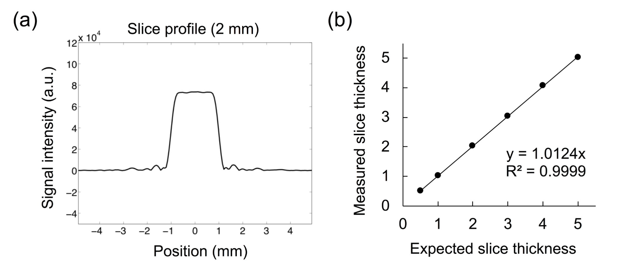
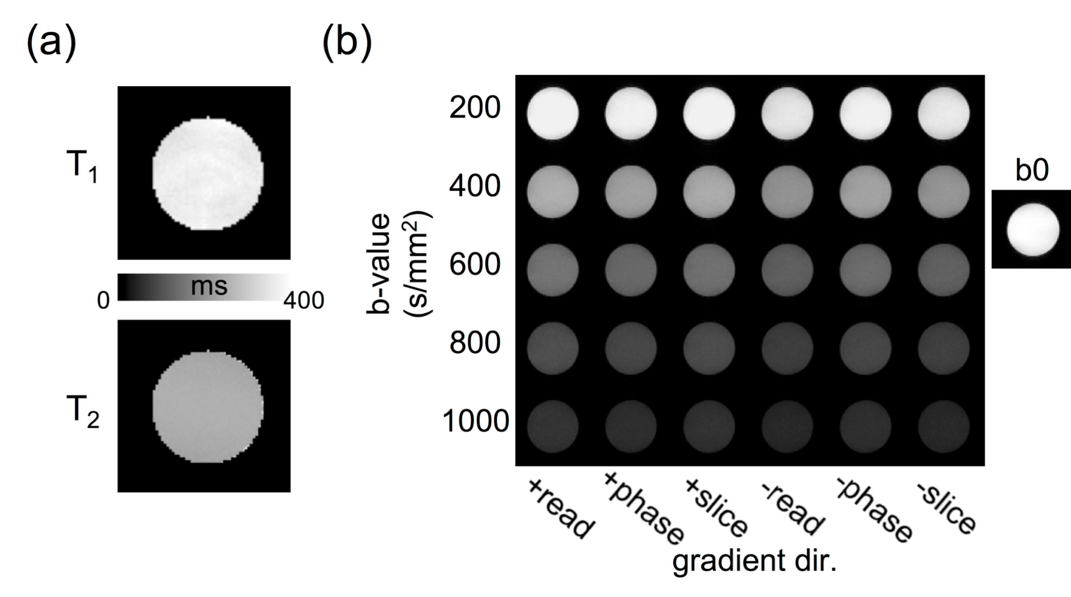
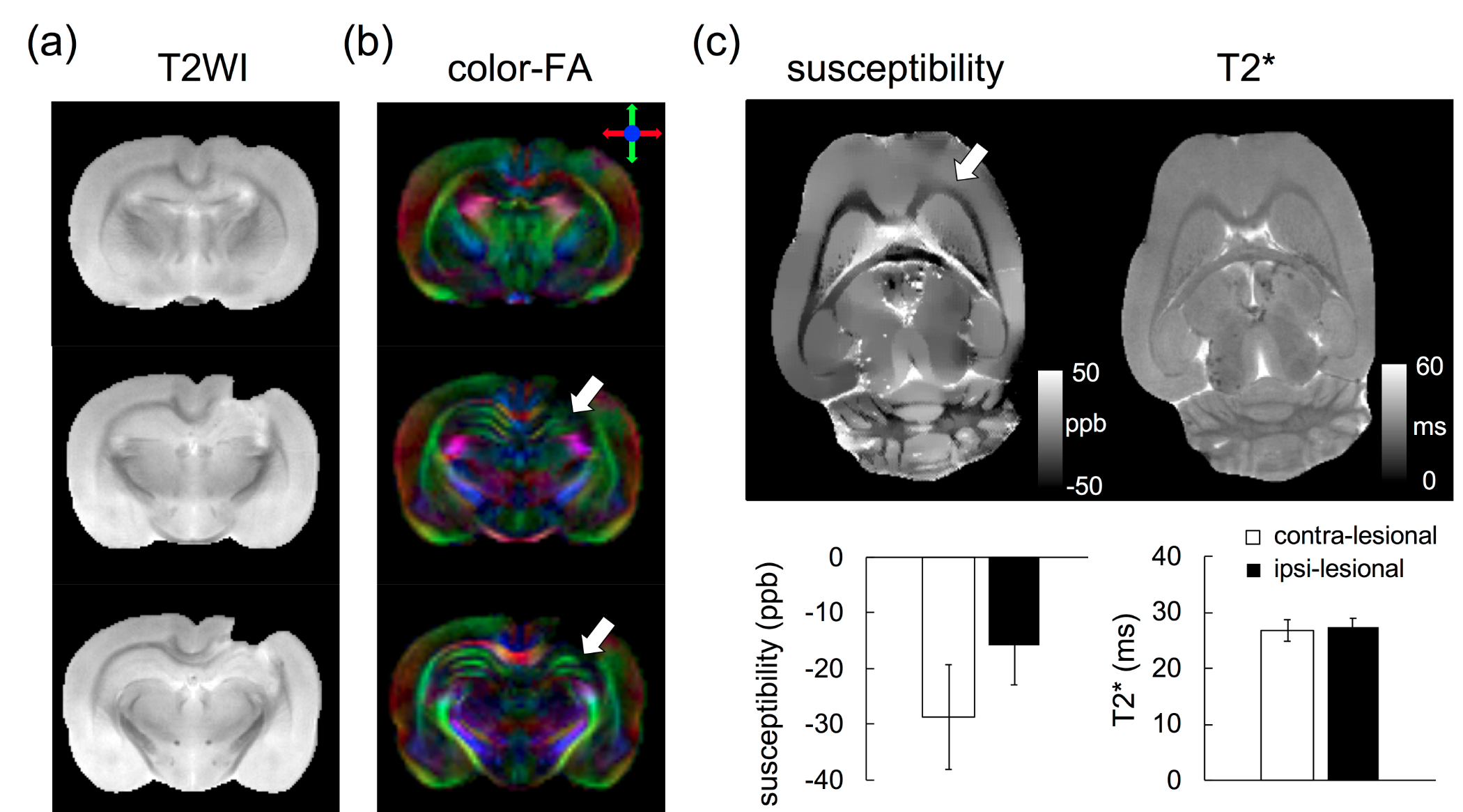
As shown in Figure 2, the slice profile measurement demonstrated an excellent agreement between the expected and measured values. Quantitative measurements on CuSO4 phantom gave T1 of 373.3±7.2 ms and T2 of 270.6±4.0 ms. The mean diffusivity of deionized water at 19.2 °C are 2.01 ± 0.02, 1.97 ± 0.02 and 1.99 ± 0.02 (10-3 mm2/s) along readout, phase and slice directions, respectively. As shown in Figure 4, the reconstructed DTI index mappings present that reasonable SNR and image quality can be obtained on our integrated system for realizing the feasibility of conducting in-vivo experiments. Figure 5 demonstrates the ability of our system on pre-clinical application in mild traumatic brain injury (mTBI) rat model.Discussions
With careful system tuning and calibration, the correctness of gradient scale setting and RF pulse output has been verified. The measured T1/T2 and ADC in phantoms were also comparable to previous findings 8,9. While the ability of our system in in-vivo rodent imaging was demonstrated, potential for pre-clinical application was also shown by using DTI and QSM on mTBI rat model. The preliminary results showed a tendency could be obtained promisingly by comparing the quantitative indices between contra-lesional and ipsi-lesional sides. Further improvements are still needed to strengthen the capability of this integrated system and its use on animal applications. Most importantly, this study can be used as a practical guide for establishing the dedicated animal imaging platform on clinical MRI scanners and facilitating the translational studies on clinical settings.Conclusion
A high-strength gradient coil dedicated for small animal imaging has been successfully integrated on a clinically used 3T MRI magnet. The feasibility of the system has been qualitatively and quantitatively demonstrated from phantom to in-vivo rodent experiments.Acknowledgements
We thank for the funding supports from National Health Research Institutes (BN-107-PP-06 and BN-107-SP-05), Taiwan Ministry of Science and Technology (106-2221-E-400-002, 107-2911-I-400-502, 107-2321-B-400-005, and 107-2221-E-400-001), and the Taiwan Central Government S & T grant (107-1901-01-19-02). We also greatly thank Dr. Piotr Starewicz to provide generous technical supports to the integration of gradient coil system.
References
1. Pfefferbaum A, Adalsteinsson E, Sullivan EV. In vivo structural imaging of the rat brain with a 3-T clinical human scanner. J Magn Reson Imaging 2004;20(5):779-785.
2. Chen F, De Keyzer F, Wang H, Vandecaveye V, Landuyt W, Bosmans H, Hermans R, Marchal G, Ni Y. Diffusion weighted imaging in small rodents using clinical MRI scanners. Methods 2007;43(1):12-20.
3. Linn J, Schwarz F, Schichor C, Wiesmann M. Cranial MRI of small rodents using a clinical MR scanner. Methods 2007;43(1):2-11.
4. Mayer D, Zahr NM, Adalsteinsson E, Rutt B, Sullivan EV, Pfefferbaum A. In vivo fiber tracking in the rat brain on a clinical 3T MRI system using a high strength insert gradient coil. Neuroimage 2007;35(3):1077-1085.
5. Aradi M, Steier R, Bukovics P, Szalay C, Perlaki G, Orsi G, Pal J, Janszky J, Doczi T, Schwarcz A. Quantitative proton MRI and MRS of the rat brain with a 3T clinical MR scanner. J Neuroradiol 2011;38(2):90-97.
6. Almeida GS, Panek R, Hallsworth A, Webber H, Papaevangelou E, Boult JKR, Jamin Y, Chesler L, Robinson SP. Pre-clinical imaging of transgenic mouse models of neuroblastoma using a dedicated 3-element solenoid coil on a clinical 3T platform. Brit J Cancer 2017;117(6):791-800.
7. Raylman RR, Ledden P, Stolin AV, Hou B, Jaliparthi G, Martone PF. Small animal, positron emission tomography-magnetic resonance imaging system based on a clinical magnetic resonance imaging scanner: evaluation of basic imaging performance. Journal of Medical Imaging 2018;5(3).
8. Kjaer L, Thomsen C, Henriksen O, Ring P, Stubgaard M, Pedersen EJ. Evaluation of relaxation time measurements by magnetic resonance imaging. A phantom study. Acta Radiol 1987;28(3):345-351.
9. Tofts PS, Lloyd D, Clark CA, Barker GJ, Parker GJ, McConville P, Baldock C, Pope JM. Test liquids for quantitative MRI measurements of self-diffusion coefficient in vivo. Magn Reson Med 2000;43(3):368-374.
Figures