1591
3D Printed Mouse Brain Holders for High Throughput Ex Vivo MRI1Department of Biomedical Engineering, Vanderbilt University, Nashville, TN, United States, 2Vanderbilt University Institute of Imaging Sciences, Vanderbilt University, Nashville, TN, United States, 3Department of Electrical Engineering, Vanderbilt University, Nashville, TN, United States
Synopsis
High throughput MRI of ex vivo mouse brains has been established as a powerful tool for studying mouse models of neurological disorders. These studies have used a RF coil array to image up to 16 mouse brains simultaneously; however, most pre-clinical MRI facilities are not equipped with multiple receivers and may not have a high field magnet with sufficient bore size to accommodate multiple coils. Here, we present designs for 3D-printed multiple mouse brain holders that can be produced inexpensively and enable high quality multiple mouse brain MRI with RF coils ≥ 25 mm diameter.
Purpose
High throughput MRI of ex vivo mouse brains has been established as a powerful tool for studying mouse models of neurological disorders 1,2. These studies have used a RF coil array to image up to 16 mouse brains simultaneously; however, most pre-clinical MRI facilities are not equipped with multiple receivers and may not have a high field magnet with sufficient bore size to accommodate multiple coils. An alternative approach that is achievable on any system is to simply load multiple brains within a standard volume coil. In order to do this in a fast, reproducible manner, a dedicated sample holder is necessary. Here, we present designs for 3D-printed multiple mouse brain holders that can be produced inexpensively and enable high quality multiple mouse brain MRI with RF coils ≥ 25 mm diameter.Materials and Methods
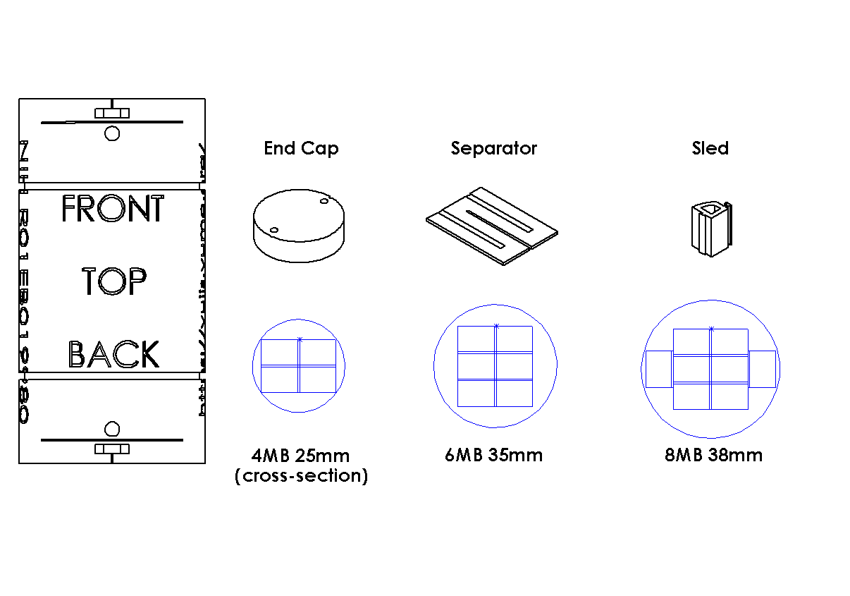
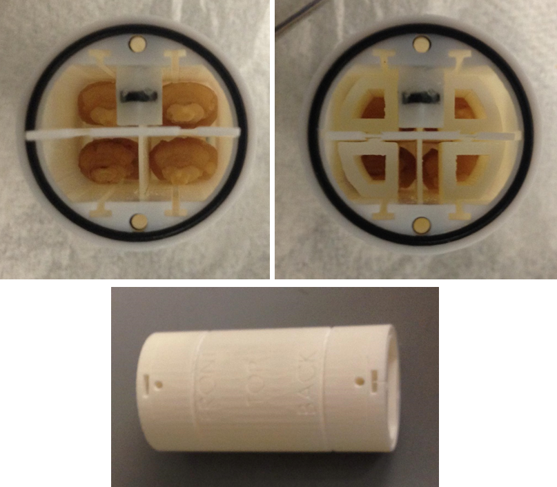
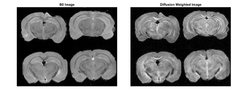
All designs were modelled in SolidWorks 2017® (Fig 1). The cross section of a mouse brain holder is a circle with an inscribed rectangular grid composed of smaller individual rectangular compartments (Fig 1). Rows and columns of compartments in the grid are separated by a thin wall to prevent physical contact between brains. The number of compartments in the grid can be determined with the following equations:$$n=floor(\frac{\sqrt{2}r}{h})$$$$m=floor(\frac{\sqrt{2}r}{w})$$Where r is the radius of the holder, h is the height of each compartment plus the distance of separation between rows, w is the width of each compartment plus the distance of separation between columns, n is the number of rows and m is the number of columns in the grid. For ex vivo mouse brain imaging, individual compartments of h x w = 7.5 x 10.5 mm2 were found suitable for a wide range of mouse different mouse strains/models. For RF coils with diameter ≥ 38 mm, the perimeter of the n x m grid of compartments can accommodate additional compartments, as seen in Fig 1. A four brain holder was fabricated to fit into a 25mm ID Doty RF litzcage coil (Doty Scientific, Columbia, SC) for use in a 7T 16-cm bore magnetic operated by a Bruker Biospec console (Billerica, MA, USA). Parts were printed in Ultra High Definition (UHD) mode in 3D Systems’ Projet 3500 HDMax with VisiJet M3 Crystal for the base material. After printing, parts were transferred to an oven heated to 62°C and left overnight to sufficiently melt off support material. Residual wax adhered to finer features in the holder were removed by placing parts in a mineral oil bath for ~1 hour in an ultrasonic cleaner. Parts were then rinsed in soap water and placed in a glass jar of hexane overnight to wash off remaining mineral oil. A set of low-scratch nylon bristles dipped in hexane were used to thoroughly scrub through small internal features (i.e. screw holes). To discern orientation, a reference marker filled with 25µM manganese chloride (MnCl2) solution was placed in a capillary tube cut to ~15mm in length then sealed and attached to the upper left corner in the interior of the holder with a film of silicone. Four chemically fixed C57BL/6J mouse brains were placed in the holder, which was filled with Fomblin Y LVAC (Fig 2) and sealed with a thin layer of silicone between the end caps and body of the holder as a measure of precaution. Three dimensional diffusion-weighted fast spin-echo MRI were acquired at 150µm isotropic resolution (Fig 3).Results and Conclusions
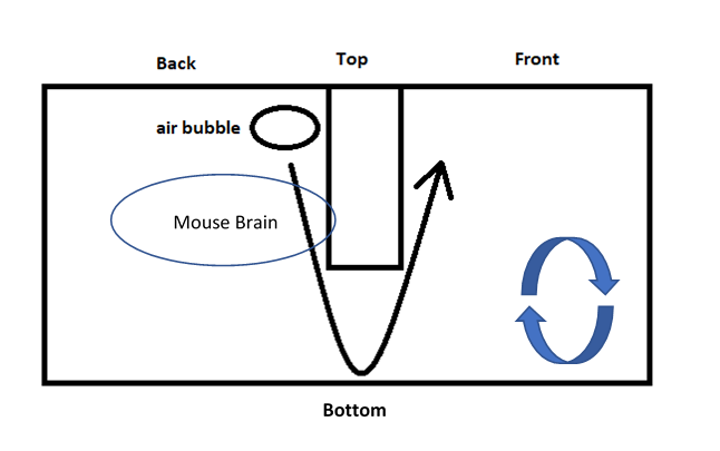
Samples were loaded within minutes and scans could start immediately following shimming and RF calibration, without need to plan imaging geometry. The resulting images were visually clean with no artifacts in the background. Brains were evenly distributed about the center such that four distinct datasets could be obtained through automated splitting of the full dataset along the midlines. No leakage was observed before and after the imaging experiment. Exterior air bubbles were effectively trapped away from the brains at one end of the holder such that the images remained unaffected (Fig 4). The usage of 3D printed mouse brain holders also serves as a proof of concept for potential designs of holders for other animal tissues. Diameters for a 4, 6, and 8 mouse brain holder for RF coil ID 25mm, 35mm, and 38mm, respectively, were shown to be easily modifiable in corresponding SolidWorks part files (.sldprt) without affecting other features.Acknowledgements
The authors thank Dr. Nicholas M. Adams and Ken Wilkens (Vanderbilt University) for assistance with 3D printing. This work is supported by the National Institutes of Health through grant number R01EB019980.References
1. Ellegood J, et al. Clustering autism – using neuroanatomical differences in 26 mouse models to gain insight into the heterogeneity. Mol Psychiatry. 2015;20(1):118-125.
2. Dazai J, et al. Multiple mouse biological loading and monitoring system for MRI. Magnetic Resonance in Medicine. 2004;52:709-715.
Figures