1570
Bilateral Breast coil for Fast Field-Cycling Relaxometric MRI1Biomedical Physics, University of Aberdeen, Aberdeen, United Kingdom, 2Aberdeen Biomedical Imaging Centre, University of Aberdeen, Aberdeen, United Kingdom
Synopsis
Our research group is investigating the use of Fast Field-Cycling MRI (FFC-MRI) for clinical applications. Recent results have confirmed the presence of interesting FFC-MRI biomarkers in breast cancer that could lead to important applications. To study this we are developing an FFC-MRI compatible breast coil for use on patients. This work is presented here and shows excellent results, paving the way to clinical applications.
Introduction
Conventional clinical MRI of the breast requires the use of exogenous contrast agents. Furthermore, the identification of lesions in dense breast tissue,with strong background enhancement, presents further challenges.
Following the work of Koenig and Brown1 our group has observed the potential of novel Fast Field-Cycling (FFC) NMR biomarkers, which do not require the introduction of contrast agents, correlating with the Nottingham Prognostic index in excised breast tumour tissue2. Significant differences were observed in the dispersion curves between healthy, malignant and peritumoural tissue that are not degraded with breast density.
We now intend to take the work forward by imaging the breasts of healthy volunteers and, later, of patients with breast cancer, using FFC-MRI3. In FFC-NMR relaxometry the magnetic field is cycled between a high polarisation field and a chosen evolution field before returning to a fixed high detection field over the course of a pulse sequence, allowing the relaxation rate (R1 =1/T1) to be determined over a wide range of field strengths. This requires tailor-made coils so the first step towards this goal, presented here, is the design, construction and testing of radiofrequency coils tailored for use with our existing 0.2-tesla, whole-body FFC-MRI scanner4.
Methods
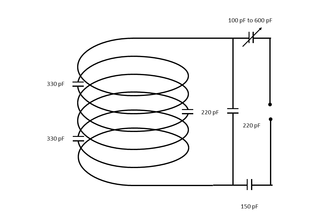
The initial coil, for proof of concept, is a unilateral device. 6 turns of 3 mm copper wire were placed on a Perspex tube of 150 mm o.d., 140 mm i.d. and 80 mm length. The circuit for the coil is shown in figure 1. The coil was enclosed in a Foamalux (Irpen Ltd., UK) box, adjacent to a secondary “dummy” Perspex tube, with their axes separated by 170 mm, to accommodate volunteers and patients with comfort.
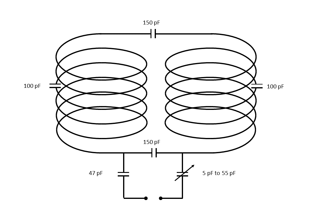
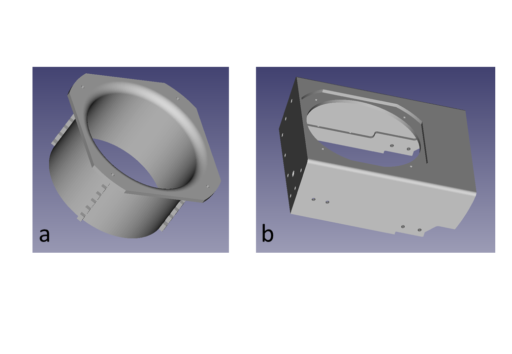
Following encouraging performance of the unilateral coil a bilateral coil, for increased versatility, was constructed. The circuit for the coil is shown in figure 2. Six turns of 3 mm diameter copper wire were wound onto each of two cylindrical supports, shown in figure 3a, produced from a design generated in Freecad by an Ultimaker-3 Extended 3-D printer (Ultimaker, Netherlands). The material used was Ultimaker Tough PLA (poly-lactic acid). The supports have an i.d. of 140 mm, an o.d. of 150 mm and a height of 90 mm. the upper 10 mm of the supports have a 10 mm radius flare to enhance comfort.
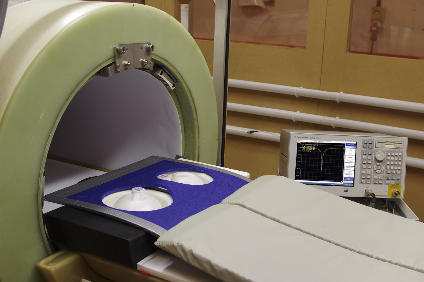
The two coils are mounted in a coil box designed in Freecad and produced in two identical halves as shown in figure 3b, by an Ultimaker 5s 3-D printer. Figure 4 shows the bilateral coil undergoing tuning and matching prior to an imaging test with a pair of 750 ml 0.8 mM MnCl2 phantoms. Images were acquired using a field-cycled spin-echo sequence with 32 ms echo time and 300 ms polarisation at 0.2 T.
Results
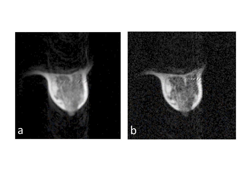
The unilateral coil has produced high quality FFC-MRI images of the left breast of a healthy volunteer. In Figure 5 contrast differences are seen between images acquired with an evolution field of 200 mT (a) and of 1 mT (b). Polarisation at 200 mT for 200 ms was followed by evolution fields for 26 ms before a spin echo images (TE = 40 ms) were acquired at 0.188 T (8 MHz). The 128 by 128 pixel images have a slice thickness was 10 mm and the field of view was 300 mm.
The bilateral coil also has a resonance frequency of 8 MHz and a Q factor, when loaded with 0.8 mM MnCl2 phantoms, of 300, which is a significant improvement on the Q factor of 160 for the unilateral coil.
Conclusion
The results obtained show that the unilateral and bilateral coils perform well and are ready for use with human studies. We intend to start clinical trials of FFC-MRI on breast cancer patients, using the bilateral coil, during 2019.Acknowledgements
This project has received funding from The Wellcome Trust Institutional Strategic Support Fund.References
1. Koenig S.H. and Brown R.D., Field-cycling relaxometry of protein solutions and tissue: Implications for MRI, Prog. Nucl. Magn. Reson. Spectrosc. 1990;22(6):487-567
2. Broche L.M. et al., Potential applications of NMR relaxometry in Medicine, 1st Working Group meeting of the European Network on Relaxometry 16 February 2017
3. Lurie D.J. et al., Fast field-cycling magnetic resonance imaging: CR Physique. 2010:11:136-148
4. Ross P.J. et al., A new human-scale fast-field cycling MRI system for clinical applications: Proc. 25th ISMRM 2017;2677
Figures