1548
A light-weight, flexible head and neck coil design for a patient-friendly MR-only Radiation Therapy workflow1GE Healthcare, Munich, Germany, 2GE Healthcare, Waukesha, WI, United States, 3GE Healthcare, Stockholm, Sweden, 4GE Global Research, Bangalore, India, 5GE Healthcare, Rochester, MN, United States, 6NeoCoil, Pewaukee, WI, United States
Synopsis
MRI is known to provide a superior soft tissue contrast when compared to CT. MR simulation offers the potential of improving target and organ at risk delineation and is therefore playing an increasingly important role in the Radiation Therapy (RT) planning workflow. Here a lightweight, highly flexible, novel coil prototype for head and neck is presented, demonstrating that a patient friendly MR-only simulation workflow for standard MR imaging and pseudo CT conversion is feasible in a clinical setting and compatible with RT fixation devices.
Introduction:
MR-only simulation for tumor delineation and dose calculation for Radiation Therapy (RT) planning is very appealing as it has the potential of improving tumor targeting, while simplifying the workflow by using a single imaging system. Silent Zero Echo Time (ZTE) MR imaging was recently demonstrated suitable for both Attenuation Correction in PET/MR and for pseudo CT conversion for Head and Neck applications1. To allow patient positioning in the MRI with the RT fixation devices and reach the desired coverage and image quality, current clinical practice often results in a very bulky and uncomfortable set-up made of a composition of different coils. Here we present a novel coil prototype specifically designed for RT using lightweight GE AIR technology2,3. This highly flexible AIR coil is easy to handle and well adapts to patient position and size and is suitable for standard MR imaging as well as for ZTE based pseudo CT image conversion.Methods:
A 3.0T GE SIGNA MR scanner (GE Healthcare, Chicago, IL) and
a prototype AIR coil were used for data acquisition on four volunteers. The
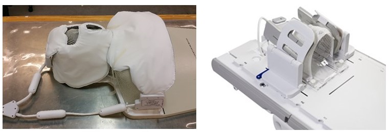
coil, depicted on the left side of Fig. 1, consists of 22 channels of which 15
located in the face, allowing for 3 different coil modes: head only, head and
neck and chest only. For signal to noise ratio (SNR) and coil coverage
assessment, phantom and volunteer scans were also performed using a reference GEM
RT open Head and Neck suite coil as shown on the right side of Fig. 1. ZTE data
were processed with a Deep Learning (DL) method where a 3D convolutional neural
network of the U-net architecture with 8-layers, Adam optimizer and RMSE cost
function was adapted to perform pseudo CT computation via image regression.
This method was trained on N=50 patients using matched pairs of ZTE and CT patient
data sets using standard product surface coils4. Results and discussion:
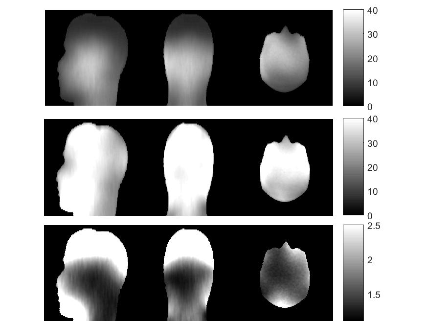
In Fig. 2 the SNR ratio of the prototype coil over the RT suite is shown. An improvement of ≥20% in SNR is measured over the whole head phantom (bottom slice of figure1). An improvement higher than a factor of two is measured on the anterior top side of the phantom.
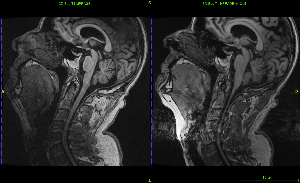
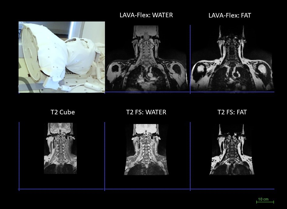
In Fig. 3 a comparison of T1 acquisition with GEM RT (left) and with the AIR coil (right) is show. In Fig. 4 coronal views of fat and water from LAVA-Flex and T2 FS sequences as well as T2 CUBE are shown for the extended coverage around the neck region using the Head and Neck coil mode of the prototype.
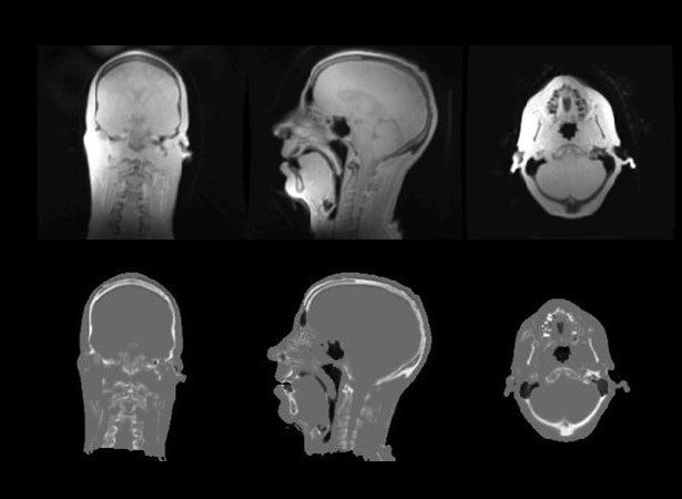
In Figure 5, ZTE images of 1.5 mm isotropic resolution are shown. At the bottom, the DL pseudo CT converted data are shown. The skull and the spine are correctly captured, while a proper training on data acquired with the prototype coil with matched resolution is expected to improve results in the neck and in the sinus region.
Conclusion:
A patient-friendly workflow which is compatible with all types of fixation devices has been enabled by a new lightweight and flexible AIR coil prototype. These first results show that the coil has appropriate coverage and image quality for standard MR imaging and silent, ZTE based pseudo CT conversion, paving the way for an optimized workflow to improve patient care with an MR-only Radiation Therapy planning workflow.Acknowledgements
No acknowledgement found.References
1. F. Wiesinger et al, ‘Zero TE‐based pseudo‐CT image conversion in the head and its application in PET/MR attenuation correction and MR‐guided radiation therapy planning’, Magn Reson Med. 2018 Oct;80(4):1440-1451.
2. S.S. Vasanawala et al., 2017. Development and Clinical Implementation of Next Generation Very Light Weight and Extremely Flexible Receiver Arrays for Pediatric MRI. arXiv preprint arXiv:1705.00224.
3. K. P McGee et al., Characterization and evaluation of a flexible MRI receive coil array for radiation therapy MR treatment planning using highly decoupled RF circuits, 2018 Phys. Med. Biol. 63 08NT02
4. S. Kaushik et al., Deep Learning based pseudo-CT computation and its application for PET/MR attenuation correction and MR-guided radiation therapy planning, ISMRM 2018
Figures