1538
Restraint system with integrated receive array for minimizing head motion during awake marmoset imaging1Centre for Functional and Metabolic Mapping, The University of Western Ontario, London, ON, Canada
Synopsis
Marmoset monkeys are an increasingly popular animal model for functional MRI studies due to their close homology with humans. To negate the confounds of anesthesia on brain activation, marmosets can be imaged awake. A restraint chair with an integrated receive array is described for minimizing motion during awake imaging. Motion was limited to 129 μm and 0.41°, allowing for comparable temporal SNR with respect to anesthetized imaging.
Introduction
The marmoset monkey is becoming an increasingly prevalent animal model in translational neuroscience studies1. By imaging animals awake, the confounding effects of anesthesia on the BOLD signal can be avoided2. Awake imaging, however, has technical challenges, as the animal must be sufficiently restrained to prevent head motion during scanning, while restraining the animal must be an efficient process to limit stress to the animal. At present, relatively few examples exist of RF-coil systems intended for awake marmoset imaging3,4. In this study, a restraint system with an integrated receive coil is described that is capable of minimizing motion during functional MRI at 9.4T, while achieving high temporal SNR. The first images acquired of an awake marmoset with this restraint system/coil are presented.Methods
The marmoset was head-fixed by clamping an implanted chamber5 to a restraint system. The restraint system was comprised of a tube for containing the body, a neck restraint, two clamps that could pivot to clamp the marmoset’s chamber, and an adjustable feeding tube and camera (Figure 1). By securing the marmoset by a chamber adhered to the skull, the design allows for future studies combining electrophysiological recordings.
The marmoset was inserted at the rear of the tube; a neck plate and rear restraint plate were then attached to confine the animal. Two side clamps were subsequently moved into place, with a screw on one clamp tightened to securely clamp the chamber. The coil former was attached to the pivoting clamps, so the coil location would be reproducible without any intervention by the handler. The coil former was designed to minimize obstructions to the visual angle of the marmoset.
The receive array (Figure 2) was comprised of five loop elements integrated into the coil former. When tightening the screw to secure the chamber, two conducting posts on one clamp push into two conductive pads on the opposite clamp to electrically connect the element circumscribing the chamber; this provides whole-brain receive sensitivity despite the presence of a chamber. RF transmission was accomplished with a 12-cm-diameter quadrature birdcage coil. Imaging was performed on a Bruker AV3HD console interfaced to a custom 15-cm-diameter gradient coil in a Magnex 9.4-T 31-cm-diameter bore magnet.
Temporal SNR was evaluated on a marmoset, when anesthetized (four 600-volume, 15-minute EPI time series) and when awake (a single 600-volume time series). Head motion during these time series was assessed by registering each volume to the central time-point volume of the respective series using AFNI6. The maximum displacement and rotation of each run was determined—multiple time series were averaged in the case of the anesthetized marmoset.
Results
The mean and maximum S12 between receive elements was -18 dB and -12 dB, respectively. Preamplifier decoupling added a further 10 – 13 dB of isolation, resulting in a mean noise correlation of 19% in vivo. The range in noise level between receive channels was 11%.
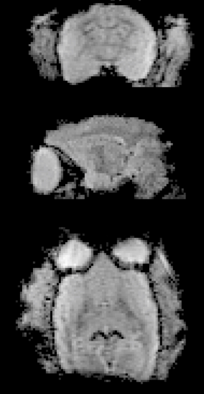
Although the chamber placed restrictions on the geometry of the receive coil, five elements could still be placed around the head for whole-brain coverage. An acceleration of two-fold was still possible in the anterior-posterior and left-right directions to reduce the echo train length (and therefore image distortion at 9.4 T). Figure 3 demonstrates the image quality in a single-volume echo-planar image accelerated two-fold in the anterior-posterior direction.
Head motion in the anesthetized marmoset was limited to an average maximum displacement of 221 μm and an average maximum rotation of 0.22°. When awake, the maximum displacement was 129 μm and the maximum rotation was 0.41° (Figure 4). Head motion had a higher variance volume-to-volume when the marmoset was awake, yet gross motion over the time series was similar to the anesthetized state—in both cases, motion was limited to less than a pixel. Minimal motion allowed the temporal SNR of the awake marmoset to be similar to that when anesthetized. Temporal SNR maps are shown in Figure 5.
Discussion
The restraint system proved to be an efficient and practical solution
for securing an awake marmoset and positioning a receive array. The marmoset
could be safely restrained, with the receive array in place,
within minutes, thereby limiting the stress to the animal. The rigidity of
the head restraint minimized motion in awake imaging, resulting in the
conservation of temporal SNR when compared to anesthetized imaging.Acknowledgements
No acknowledgement found.References
- Okano H, Hikishima K, Iriki A, Sasaki E. The common marmoset as a novel animal model system for biomedical and neuroscience research applications. Seminars in Fetal & Neonatal Medicine 2012;17(6):336-340.
- Vanduffel W, Fize D, Mandeville JB, Nelissen K, Van Hecke P, Rosen BR, Tootell RBH, Orban GA. Visual motion processing investigated using contrast agent-enhanced fMRI in awake behaving monkeys. Neuron 2001;32(4):565-577.
- Papoti D, Yen CC, Hung CC, Ciuchta J, Leopold DA, Silva AC. Design and implementation of embedded 8-channel receive-only arrays for whole-brain MRI and fMRI of conscious awake marmosets. Magn Reson Med 2017;78(1):387-398.
- Mareyam A, Sharma J, Desai M, Sander CY, Frederick E, Bartelle BB, Takahashi A, Wald LL. Marmoset coil arrays for functional imaging of awake monkeys at 9.4T and 3T. Proc Intl Soc Mag Reson Med 26 (2018):4280.
- Johnston KD, Barker K, Schaeffer L, Schaeffer D, Everling S. Methods for chair restraint and training of the common marmoset on oculomotor tasks. Journal of Neurophysiology 2018;119:1636–1646.
- Cox RW. AFNI: Software for analysis and visualization of functional magnetic resonance neuroimages. Comput Biomed Res 1996;29(3):162-173.
Figures