1526
A 16-Channel Array Coil for Anesthetized Monkey Multi-modal Neuroimaging at 3T1Interdisciplinary Institute of Neuroscience and Technology, Qiushi Academy for Advanced Studies, College of Biomedical Engineering & Instrument Science, Zhejiang University, Hangzhou, China, 2Oregon National Primate Research Center, Oregon Health & Science University, Beaverton, OR, United States
Synopsis
A 16-channel array coil has been developed in our laboratory for anesthetized monkey brain imaging at 3T, with a specialized design to accommodate multi-modal devices. Its performance has been evaluated on an anesthetized macaque. Compared to the commercially-available pediatric coil, the 16-channel monkey head coil showed improved receive sensitivity and superior acceleration performance. Further, the presented coil can accommodate multi-modal devices, allowing simultaneous optical imaging, neural recording, and stimulation, during high-field MRI studies. It is hoped that the proposed array coil could benefit a broad scope of research in frontier neuroscience.
Introduction
Non-human-primates have provided valuable animal models in frontier neuroscience research. Multi-modal neuroimaging and neuroengineering through multiple methods, e.g., MRI, optical imaging, and electrophysiology for information collection, and optogenetics, ultrasound, and infrared light for neural stimulation could offer valuable opportunities to explore the brain connectome through different spatial/temporal scales and dimensions [1-2]. High-field (≥1.5T) MRI has shown its advantages in the noninvasive visualization of brain anatomy and functionality with increased sensitivity for contrast mechanisms and substantial gains in spatial resolution. However, until now, few animal RF coils have been developed to be compatible with multi-modal imaging/stimulation approaches. To address this problem, a 16-channel array coil has been developed in our laboratory for anesthetized monkey brain imaging at 3T, with a specialized design to accommodate multi-modal devices. Its performance has been evaluated on an anesthetized macaque.Method
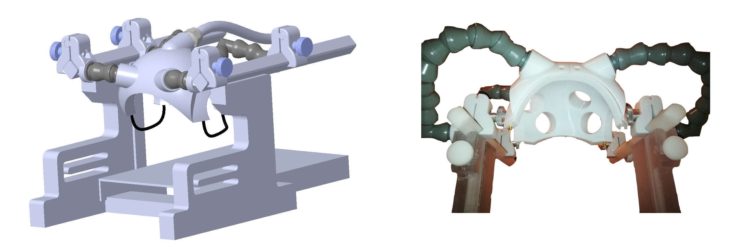
The coil consists of sixteen 4cm-diameter receive-only loops tuned to 123.23 MHz in an overlapping array configuration. Each coil element contains active detuning and pre-amp decoupling circuitry. The preamplifier was kept 50cm away from each coil element to leave space for head-fixation and multi-modal imaging devices. A lattice balun was used at the drive point to each element to eliminate cable common-mode current issues. As shown in Fig.1, the coil was constructed on a 3D-printed former made from polycarbonate using the Fused Deposition Modelling (FDM) (Virtumed LLC, Minneapolis, MN, USA). Mounting was done using the loc-line 0.75-inch modular hose to allow adjustable positioning of the coil. Two receive loops near the animal’s ears are removable for easy installation of ear-bars. Multi-modal imaging capabilities were enabled through placing four openings in the housing to allow direct access of brain regions such as temporal, parietal and occipital lobes.
All measurements were performed on a 3T clinical scanner (Prisma, Siemens, Erlangen, Germany) equipped with a whole-body gradient set (80mT/m and 200T/m/s). Images were acquired of an anesthetized macaque (male, 9.6 kg). It was placed in sphinx position inside the MRI bore when using the 16ch monkey coil described here, and in the supine position when using a 16-channel pediatric coil provided by the manufacturer4. The macaque was anesthetized with 1.4%-2% isoflurane, sufentanyl (3ug/kg), and vecuronium (0.25mg/kg), and maintained with end-tidal 0.3% isoflurane in air, sufentanyl (2ug/kg*h) and vecuronium (0.1mg/kg*h). All procedures were in accordance with NIH standards and approved by the local Institutional Animal Care Committee.
Noise correlation was estimated from thermal noise data acquired without RF excitation, SNR and g-factor maps were obtained from PD-weighted FLASH images (TR/TE/α: 10ms/4.8ms/15°, 1×1×5mm3) [3]. Diffusion-weighted images [TR/TE: 6,700ms/73ms, 1mm isotropic, GRAPPA 2, b-values: 0/1,000, scan time: 28’30”] were also acquired to calculate the fractional anisotropy (FA). Imaging parameters were kept the same when using the pediatric and monkey coils.
Results
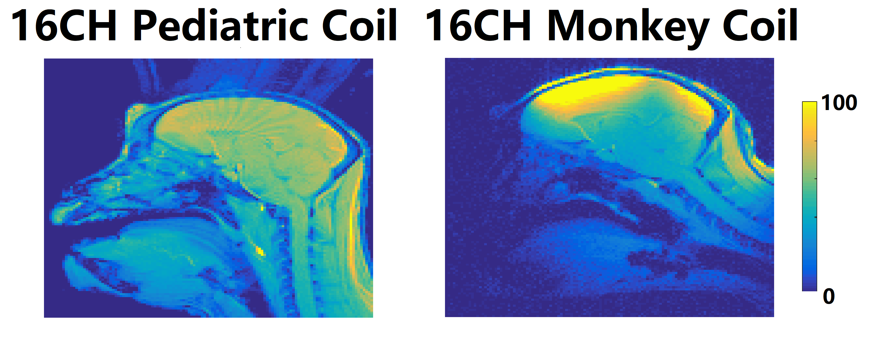
Fig.2 shows that compared to the pediatric coil, the custom-built 16-channel monkey coil has 2-fold SNR improvement in cortical regions, and only slightly lower SNR in deep brain areas, which may be attributed to the small-diameter loop elements of the custom-built coil.
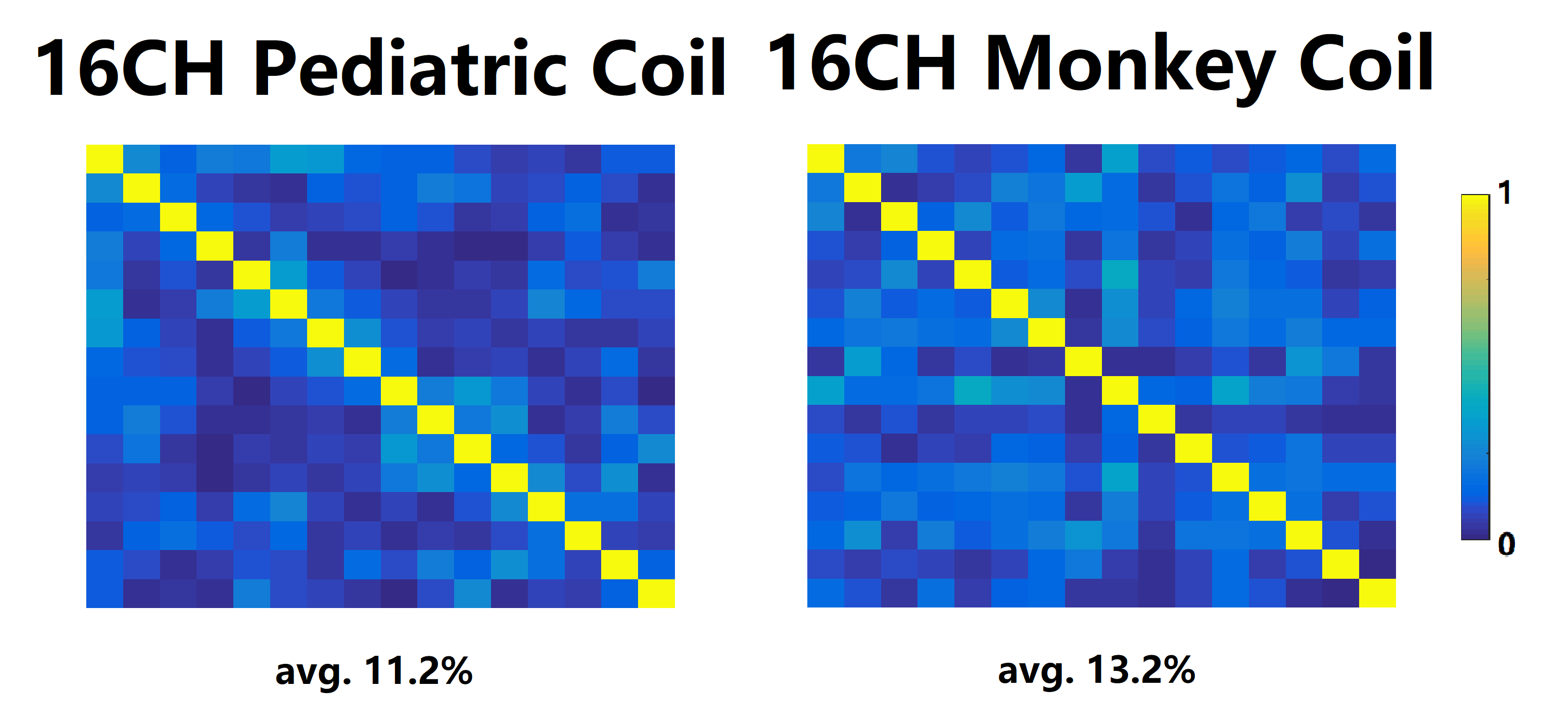
Fig.3 shows noise correlation of the monkey head array ranging from 1.4% to 39.7% (avg. 13.2%), comparable to the pediatric coil (0.08% to 33.9%, avg. 11.2%), indicating all 16 loop elements have been well decoupled.
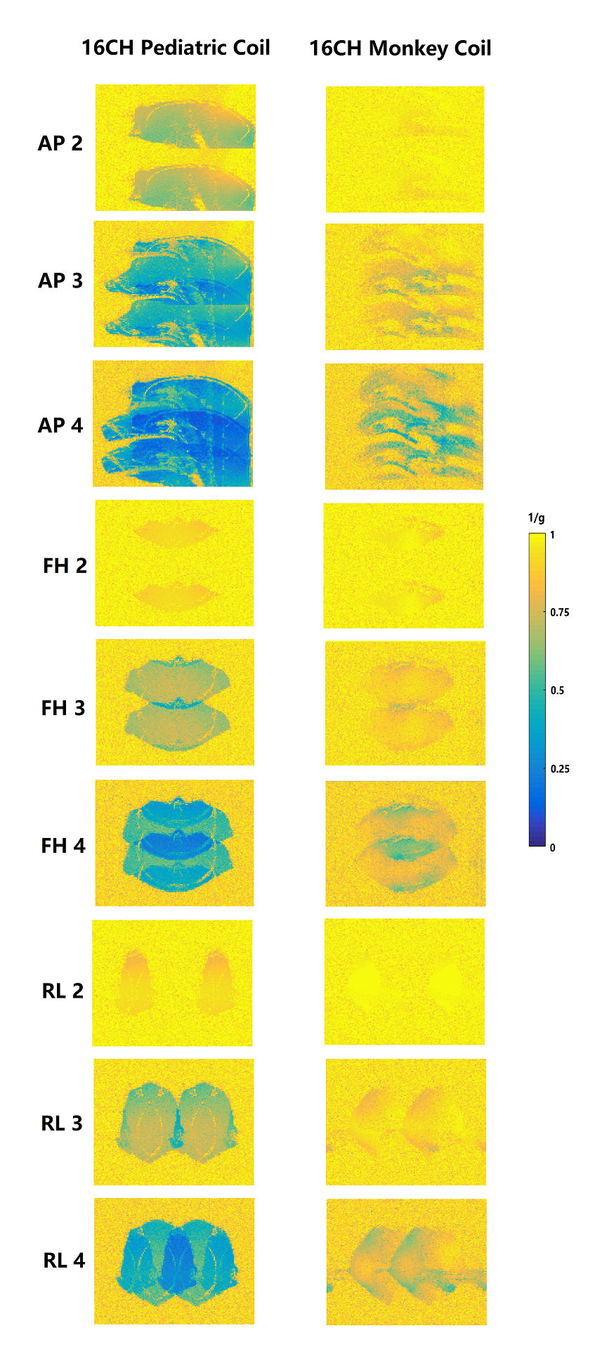
Fig.4 shows the 1/g-factor obtained for three orthogonal phase encoding directions. The monkey head array is much higher than that of the pediatric coil, suggesting its superior acceleration performance, or less of an SNR penalty, in parallel imaging. Even for high acceleration rate (>2), a 1/g-factor of 0.8 (GRAPPA 3) and 0.7 (GRAPPA 4) could be maintained in most brain regions.
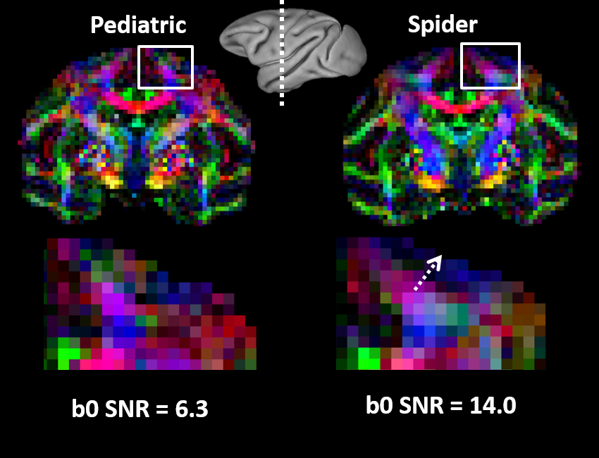
Fig.5. shows that fine structure could be well discerned with the improved SNR (14.0 for the b0 image) of 16-channel monkey coil vs commercial pediatric coil (6.3 for the b0 image).
Discussion and Conclusions
Compared to the commercially-available pediatric coil, the 16-channel monkey head coil showed improved receive sensitivity and superior acceleration performance. Further, the presented coil can accommodate multi-modal devices, allowing simultaneous optical imaging, neural recording and stimulation, during high-field MRI studies. It is hoped that the proposed array coil could benefit a broad scope of research in frontier neuroscience.Acknowledgements
National Natural Science Foundation 81701774 and 61771423, NIH NS093998. We thank Michael Reusz, Zheng Liu, and Jialu Zhang for helpful discussion and technical support.References
[1] Chernov, Mykyta, and Anna Wang Roe. "Infrared neural stimulation: a new stimulation tool for central nervous system applications." Neurophotonics 1.1 (2014): 011011.
[2] Roe, Anna Wang, et al. "In vivo mapping of cortical columnar networks in the monkey with focal electrical and optical stimulation." Frontiers in neuroanatomy 9 (2015): 135.
[3] Kellman, Peter, and Elliot R. McVeigh. "Image reconstruction in SNR units: a general method for SNR measurement." Magnetic resonance in medicine 54.6 (2005): 1439-1447.
[4] https://usa.healthcare.siemens.com/magnetic-resonance-imaging/options-and-upgrades/coils/pediatric-16
Figures