1517
Opencage Radiofrequency Coil Inspired by Metamaterials1ESPCI, Institut Langevin, PSL Research University, Paris, France, 2Direction de la Recherche Fondamentale / Institut Joliot / NeuroSpin, Commissariat à l’Energie Atomique et aux Energies Alternatives, Gif sur Yvette, France, 3Université Paris Saclay, Saclay, France
Synopsis
We propose a volume radiofrequency coil for MRI that provides a lateral access to its inner volume. This coil, called «opencage», is designed by revisiting birdcage coils as metamaterial transmission line with broken periodicity. An opencage dedicated for imaging at 7T of the head of small rodents is developed. The design of this opencage is optimized using numerical simulation. Finally in-vivo preclinical imaging of the head of a mouse is presented. We show that the opencage coil efficiency is similar, especially in terms of field homogeneity and SNR as a conventional 8-legs birdcage coil.
The concept of an opencage RF coil
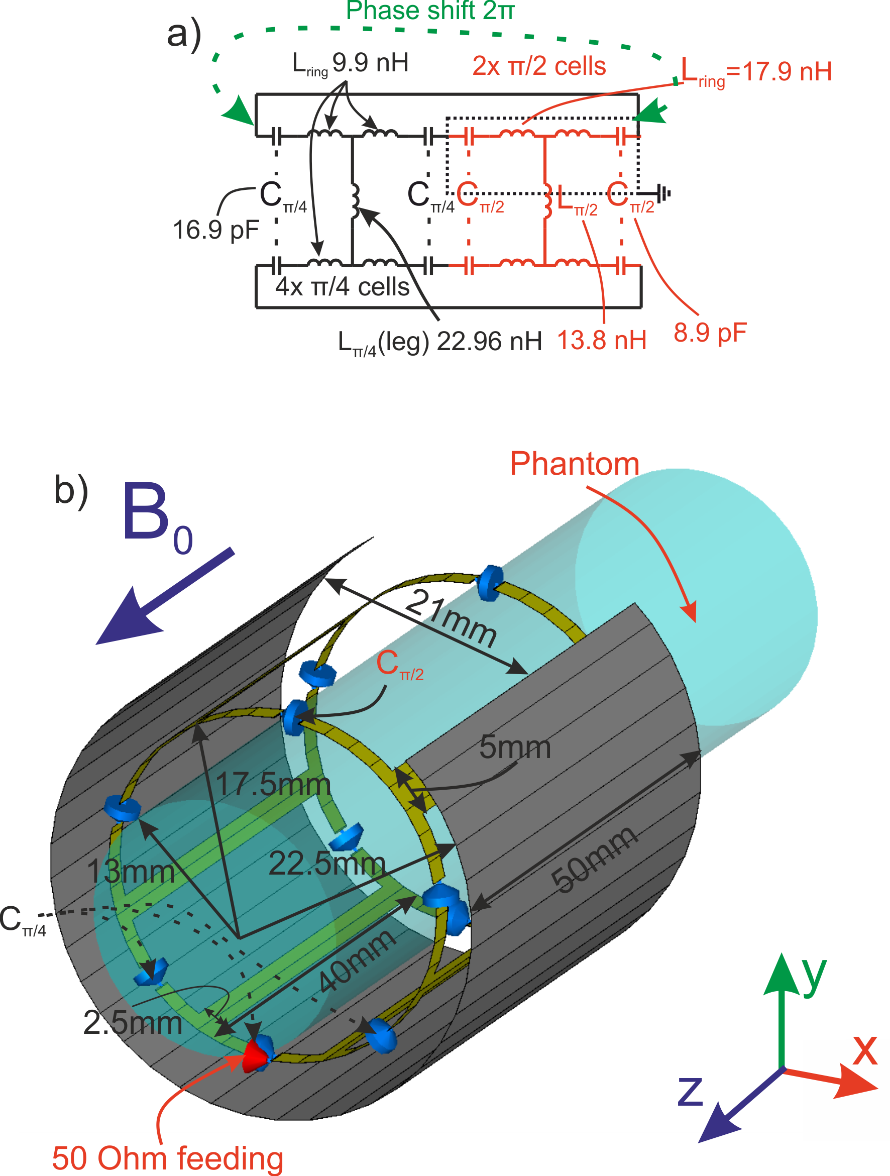
Introduced in 1985 [1] birdcage coil is now the most used volume RF coil in MRI. It is made of interconnected parallel metallic rods (legs) periodically arranged and forming a cylinder. The closer the legs and the more homogeneous B1 field. However, this geometry prevents a convenient access to the volume inside the birdcage that can be problematic for some applications especially when it is used as a head coil. One solution consists in increasing the spacing between all the legs. However the field homogeneity regresses. Here we introduce the opencage coil as a trade-off between these two contradictory aims between access and homogeneity. It requires the realization of a non-periodic structure. This can be achieved by adjusting individually the current distribution on each leg. To perform this goal one can consider a high-pass birdcage coil [1, 2] as a left-handed metamaterial. Indeed, the equivalent electrical scheme of a birdcage is a ladder network of lumped elements. One unit cell is made of a leg capacitance, a leg inductance and a ring inductance [3, 4]. To engineer the wave propagation along the transmission, the phase shift and intrinsic impedance is fixed for each unit cell of the ladder. As for birdcage, in an opencage coil, the sum of phase shifts of each unit cell should still be equal to 2π for the fundamental mode. Here two different types of unit cells are considered. By adjusting the capacitance and the inductances, these cells are designed in order to achieve respectively π/4 and π/2 phase shift with a constant impedance.Design and full-wave validation
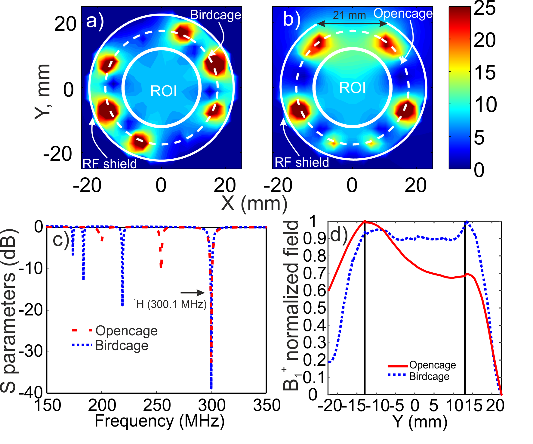
The proposed opencage coil is designed for small animal imaging at 7T. Here, four π/4 cells are connected to two π/2 cells to give form the equivalent circuit of the opencage as illustrated in Fig. 1a. Capacitor values of 16.9 pF and 8.9 pF have been used for the π/4 and the π/2 cells respectively. The desired inductances are obtained by adjusting the leg widths on the cells. Two different leg widths of 2.5mm and 5mm have been used on the π/4 and the π/2 cells, respectively. The design of the opencage coil has been validated by performing full wave numerical simulations in commercial software CST Microwave studio 2018 (Fig. 1b). The opencage coil is driven linearly by a single 50-Ohm port. The coil is loaded by a lossy homogeneous cylindrical phantom. The setup is placed inside a long metal cylinder simulating to the MRI bore. This configuration of the coil is compared to an 8-legs birdcage coil (Fig. 2a, b). All coils are matched and tuned at 300.1 MHz (Fig. 2c). The normalized profiles of B1+ field are presented in Fig. 2d. In order to estimate B1+ field homogeneity of both coils, standard deviation (SD) of the simulated field in the phantom has been calculated on the region where the brain of a mouse can be placed. For the birdcage coil SD is 0.63 µT, while for the opencage coil SD is 0.69 µT. Therefore, a similar homogeneity is obtained with this opencage while providing an easy access to the inner volume.Experimental validation of the proposed concept
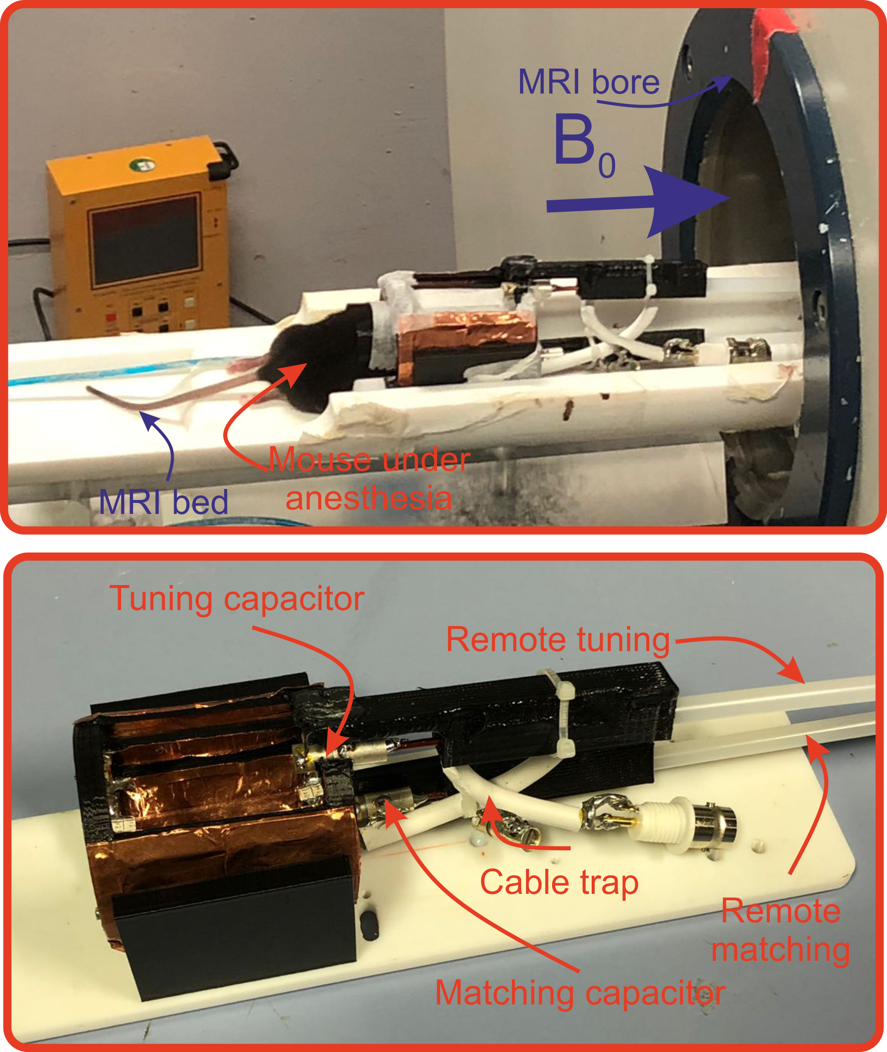
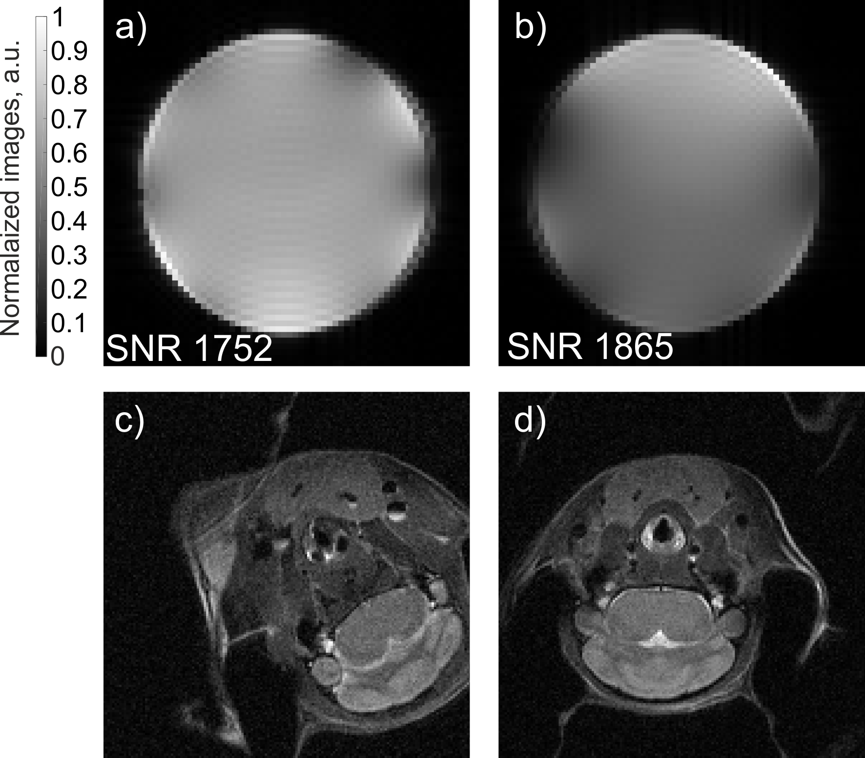
The opencage coil (Fig. 3) and the 8-legs birdcage coil are assembled in order to make an experimental comparison. The coils are tuned and matched at 300.1 MHz. Normalized images of a cylindrical Phantom (Fig. 4 a, b) have been acquired with Bruker PharmaScan 7T preclinical MRI scanner. The SNR of the images has been estimated as a mean signal divided by a standard deviation of a noise in a ROI of 10 by 10 voxels. For these images, calculated average SNR is equal to 1865 for the opencage coil and 1752 for the 8-legs birdcage coil. Therefore, SNR of two images is comparable. The two coils are tested with an anesthetized mouse (Fig. 4 c, d). The images demonstrate similar good SNR in a broad spatial FOV for both coils. This result confirms that the opencage coil can achieve a high homogeneity performance while providing a wide aperture to the inner volume.Conclusion
We have shown a practical implementation of the opencage radiofrequency coil for ultra-high field preclinical MRI. The opencage coil is birdcage-like coil providing a wide access to the inner volume. This coil can be used for many applications where the usage of a birdcage coil is not convenient due to a high density of legs. For instance, in some specific experiments using ultrasound probing, an access to the sample is required during the small animal imaging at 7T.Acknowledgements
This project has received funding from the European Union’s Horizon 2020 research and innovation program under grant agreement No 736937. This work is supported by LABEX WIFI (Laboratory of Excellence within the French Program “Investments for the Future”) under references ANR-10-LABX-24 and ANR-10-IDEX-0001-02 PSL.References
1 Cecil E. Hayes, William A. Edelstein, John F. Schenck, Otward M. Mueller, Matthew Eash, An Efficient, Highly Homogeneous Radiofrequency Coil for Whole-Body NMR Imaging at 1.5 T, Journal of Magnetic Resonance, vol. 63, p. 622-628, (1985).
2 Cecil E. Hayes, The development of the birdcage resonator: a historical perspective, NMR Biomed.,vol. 22, p. 908-918, (2009).
3 Chih-Liang Chin, Christopher M. Collins, Shizhe Li,Bernard J. Dardzinski, and Michael B. Smith, BirdcageBuilder: Design of Specified-Geometry Birdcage Coils with Desired Current Pattern and Resonant Frequency,
4 Christophe Caloz, Tatsuo Itoh, Electromagnetic Metamaterials: Transmission Line Theory and Microwave Applications, John Wiley and Sons Ltd, (2006).
Figures