1501
WEARABLE AND STRETCHABLE SURFACE BREAST COIL1Industrial PhD Program in Advanced Biomedical Technologies, Dokuz Eylul University, Izmir, Turkey, 2Institute of Biomedical Engineering, Bogazici University, Istanbul, Turkey, 3Department of Electrical and Electronics Engineering, Ege University, Izmir, Turkey
Synopsis
A novel wearable and stretchable single loop breast coil made from conductive thread is introduced with a coil geometry which can expand and retract in accordance with the breast while preserving good quality factor. Images of both the small phantom and the medium phantom could be acquired by S-M coil. Similarly, M-L breast coil was used to obtain the images of both the medium phantom and the large phantom. Highest SNR was obtained when small phantom was imaged using S-M coil and the lowest SNR was obtained with large phantom when it was imaged using M-L coil.
INTRODUCTION
Research and development of new breast coils1,2 maintain its importance owing to prevalence of breast cancer all over the world. Contrast enhanced breast MRI, having 90% of sensitivity and 75% of specificity, provides early detection of breast cancer3. In practical clinical applications, often fixed medium-sized breast coils are used in breast MRI. When the size of the patient’s breast does not fit to the breast coil, filling factor of the coil is affected, and consequently, image quality is degraded. In addition, even though frequency shift resulting from low filling factor can be somewhat compensated by image processing, images of trapped breast inside the coil may not provide sufficient diagnostic information. The requirement for a flexible coil is conspicuous considering not only the image quality and diagnostic issues, but also patient’s comfort. In this study, a novel wearable and stretchable surface breast coil made from conductive thread is introduced to overcome the inadequacies of the fixed coil size based breast MRI. Although flexible coils have been suggested, a stretchable coil using conductive textile has not been made. The proposed coil geometry can expand and retract in accordance with the breast while preserving good quality factor.METHODS
Phantoms: Three different oval shaped breast phantoms (small, medium, and large) with base diameters 100 mm, 150 mm, and 200 mm, respectively, and with heights 90 mm, 135 mm, and 180 mm, respectively, were printed in a 3D printer (Ultimaker2, Netherlands).
Stretchable Textile Coil: The superconductive silver thread (Statex, DE) was woven having a width of 4 cm via full needle rib technique, and fed with lycra elastofibre. The resultant fabric having 115.56% of stretchability was cut into two pieces of 11 cm and 12 cm. A connection circuit was designed to combine the ends of the loop coil and to enable any circuit component to be soldered as well. 11 cm of breast coil was compatible with both small and medium phantom (Figure 1), so it was called S-M breast coil. Similarly, 12 cm of breast coil was compatible with both medium and large phantom (Figure 2), therefore it was called M-L breast coil.
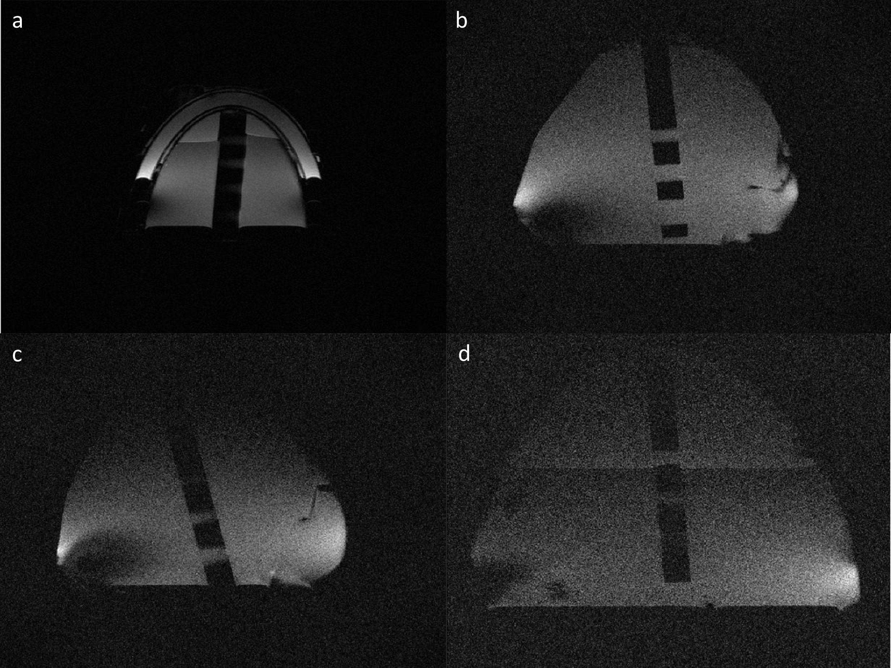
In-vitro Studies: The images of three phantoms filled with corn oil and water were acquired at 3T MRI system (Siemens, DE) using T2 TSE sequence (FoV=160mm, TR=5410 ms, TE=85 ms Slice thickness= 3 mm). The images of the small and the medium phantom were acquired using S-M breast coil, and images of the medium and the large phantom were obtained using M-L breast coil. To assess the image quality of the acquired phantom images, signal-to-noise ratio (SNR) was calculated for each image using mean pixel intensity of an ROI placed on phantom as signal and standard deviation of an ROI introduced in background as noise.
RESULTS
Matching Circuit: The stretchable textile breast coils sustained their tuned and matched state (Table 1) when they were stretched and filled with different sized phantoms.
In Vitro Studies: Calculated SNR values for each acquired image were ranging from 8.62 to 49.32. Highest SNR was obtained when small phantom was imaged using S-M coil and the lowest SNR was obtained with large phantom when it was imaged using M-L coil. Detailed information is provided in (Table 2). Inplane resolution of the scan was 0.39 mm/pixel.
DISCUSSION
The stretchable textile coil could be tuned like any other traditional coil. However, its stable impedance after tuning was independent from the size of the phantom, and was unique to itself. This characteristic enables a single matching circuit to be used even though the size of the subject changes. To test stretchable coil, a simple loop antenna was made. As expected, the small phantom had larger SNR than the large one due to decreased magnetic field as phantom size was increased. Especially in axial images of resolution plate, the effect of insufficient coil geometry was notable.
In conclusion, image quality of the acquired images were reasonable and applicable for clinical use although the geometry of the stretchable textile coil was rather simple and it was single channel. The idea of using a conductive thread to create RF coils is feasible and promising with further enhancements concerning the geometry, the number of channels, and low noise amplifier (LNA) structure.
Acknowledgements
This study was partly funded by TUBITAK (The Scientific and Technological Research Council of Turkey) under grant no. 116E155.References
1. Ileana Hancu et al, Mag Res in Med 00:00–00 (2015)
2. Anderson N. Nnewihe et al, Mag Res in Med 66:281–289 (2011)
3. S.H. Heywang-Köbrunner et al, The Breast, 22 (2013) S77-S82
Figures