1495
Towards uncompromised merging of 1H and 31P receive arrays for multi nuclear metabolic imaging in the brain at 7T1Image Sciences Institute, University Medical Center Utrecht, Utrecht, Netherlands, 2University of Twente, Enschede, Netherlands
Synopsis
In order to optimally integrate 31P MRS and 1H MRI, a 1H birdcage coil is combined with a 16 channel dual-tuned 31P/1H receive loop array for 7T, inserted in a 31P
Introduction
31P MRS can be used for studying several brain pathologies (e.g. brain tumors1, schizophrenia2) in which abnormal metabolism of phospholipids occur3. Together with MR imaging, it may become a non-invasive tool to provide a better diagnosis and monitoring of brain disease. One approach to optimally integrate 31P MRS and MRI with reduced patient manipulation and more accurate spectra localization is by using a double-tuned coil. In particular, double-tuned 31P/1H birdcage coils have proven to provide sufficient signal uniformity to facilitate 31P metabolite measurements at 1.5 and 3T4-6 but, for many applications, a better SNR than available at 3T is needed7. In this study, a new unshielded 1H birdcage coil, combined with a 16 channel dual tuned 31P/1H receive loop array for 7T was designed with the aim of maximizing detection sensitivity and transmit field homogeneity for 31P while maintaining adequate imaging performance for 1H. For a uniform phosphorous excitation, the whole-body coil developed by Loring et al.8 was used. In order to be able to use the whole-body coil for 31P transmission, the standard RF shield of the 1H birdcage coil is removed. The aim of our study was to compare the unshielded 1H transmit and 16 channel 1H/31P receive coil to the commonly used 1H birdcage coil with a 32 channel 1H receive array (NOVA head coil, NOVA medical, Wilmington, USA) on RF safety, transmit efficiency and signal-to-noise ratio (SNR), while providing maximized SNR for 31P MRS .Methods
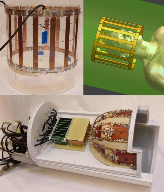
Finite difference time-domain simulations (Sim4Life, Zurich Medtech, Zurich, Switzerland) were used to evaluate the RF safety of the new unshielded 2Tx high-pass quadrature birdcage coil (Fig.1) and the NOVA birdcage coil. B1+ and 10g-averaged SAR distributions for both coils were simulated on the human models Duke (Male 34 years old), Billie (Female, 11 years old), and Thelonious (Male, 6 years old) of the virtual family9. After obtaining local IRB approval, three healthy volunteers were scanned with both coils on a 7T Achieva MR system (Philips Healthcare, Best, The Netherlands), in order to obtain B1+ (DREAM, FA=40º, TR/TE=3.7/1.4ms, FOV=224x224x224mm3, resolution=3.5x3.5x3.5mm3, 20 slices) and SNR (FA=90º, TR/TE=3000/1.7ms, FOV=350x350x25mm3, resolution=5x5x5mm3, 3 slices) maps for the 1H signal of both coils, using the same input power. 3D 31P MRSI data was obtained with the 16 channel receiver array using a 18 degree flip angle excited with the bodycoil and TR of 200ms at 2.5cm resolution with 5 hamming weighted averages in a scan time of 15 minutes.Results
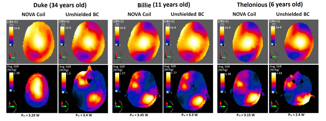
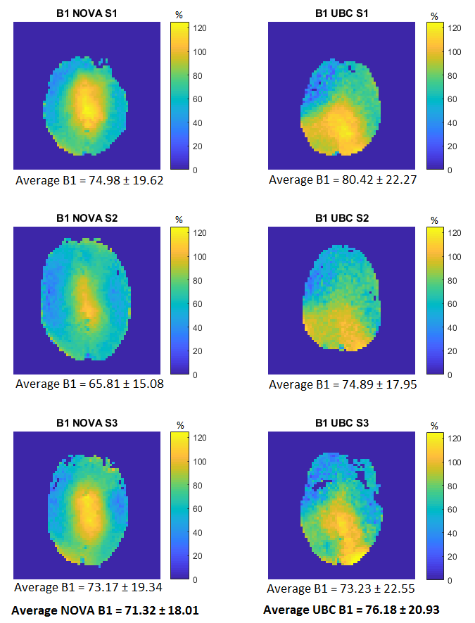
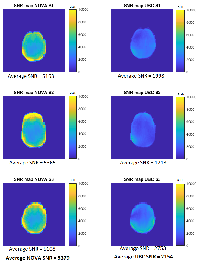
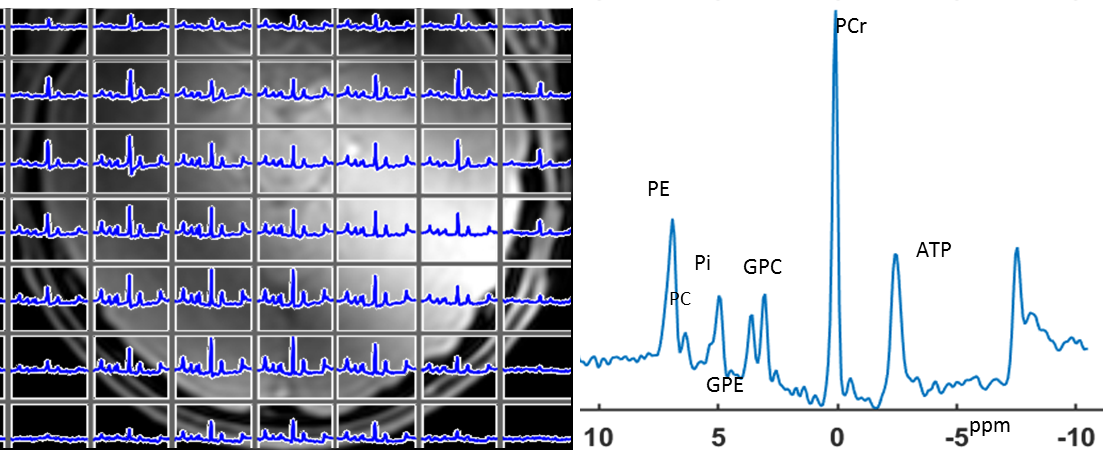
Simulation results can be found in Fig.2, where the slice with the highest 10g-averaged local SAR for a maximum B1+=1µT in the coil isocenter is displayed for each subject and coil. Similar B1+ and SAR distributions can be observed for both coils. The experimental B1+ distribution is shown in Fig.3, in which the maximum B1+ for both coils and all subjects is around 125% of the refB1+ (9µT) for the same input power. However, although the peak B1+ is less centered in the case of the unshielded birdcage, its average B1+ is 6.8% higher. The SNR maps presented in Fig.4 indicate a decrease in the slice average SNR of 60% in the unshielded double-tuned coil with respect to the NOVA coil. High SNR 31P MRSI data was obtained throughout the brain (Fig. 5).Discussion
Anatomical distinctions due to the age difference cause dissimilarities in the B1+ and SAR distributions between the simulated subjects. In the NOVA simulations, the SAR slightly decreases when the subject head is smaller because less power is required to achieve 1µT in the center. The experimental B1+ maps show a 6.8% increment of average B1+ for the unshielded birdcage as compared to the NOVA birdcage but also a higher standard deviation as a result of its higher field inhomogeneity. A decrease in SNR of the double-tuned receivers as compared to the NOVA coil can be expected as a result of lower Q-factor. However, the large decrease in signal-to-noise ratio that is observed in this case is also due to the fact that the transmit birdcage is currently not detuned during receive mode, which is a possible improvement to be included in future work. On the other hand, superb SNR for 31P MRS throughout the brain was obtained with the integrated setup.Conclusion
Based on the present study, a 31P/1H double-tuned receive array combined with an unshielded birdcage coil has an adequate 1H transmit performance, and has the potential for accelerated imaging with the 16 double-tuned receive loops. This enables optimized 31P MRS and accelerated 1H MRI acquisition at 7T.Acknowledgements
No acknowledgement found.References
References
[1] Podo, F. Tumour phospholipid metabolism. NMR in Biomedicine. 1999; 12(7), 413–439.
[2] Wijnen, J. P., Scheenen, T. W. J., Klomp, D. W. J., & Heerschap, A. P Magnetic resonance spectroscopic imaging with polarisation transfer of phosphomono- and diesters at 3 T in the human brain : relation with age and spatial differences. NMR in Biomedicine. 2010; 23(8), 968–976.
[3] Khlebnikov, V., Wijnen, J., Van der Kemp, W. J. M. & Klomp, D. W. J. Chapter Five – 31P MRSI Studies in Patients with Cancer. Annual Reports on NMr spectroscopy. 2016;87. 319-368.
[4] Matson, G. B., Vermathen, P. & Hill, T. C. A practical double-tuned1H/31P quadrature birdcage headcoil optimized for 31P operation. Magnetic Resonance in Medicine - MAGN RESON MED.1999; 42. 173-182.
[5] Derby, K., Tropp, J., & Hawryszko, C. Design and evaluation of a novel dual-tuned resonator for spectroscopic imaging. Journal of Magnetic Resonance.1990; (1969), 86(3), 645–651.
[6] Greenman, R. L., & Rakow-Penner, R. Evaluation of the RF field uniformity of a double-tuned 31P/1H birdcage RF coil for spin-echo MRI/MRS of the diabetic foot. Journal of Magnetic Resonance Imaging. 2005; 22(3), 427–432.
[7] Moser, E. Ultra-high-field magnetic resonance: Why and when? World Journal of Radiology.2010; 2(1), 37.
[8] Löring, J., van der Kemp, W. J. M., Almujayyaz, S., van Oorschot, J. W. M., Luijten, P. R., & Klomp, D. W. J. Whole-body radiofrequency coil for31P MRSI at 7T. NMR in Biomedicine. 2016; 29(6), 709–720.
[9] Christ, A., Kainz, W., Hahn, E. G., et al. The Virtual Family - Development of surface-based anatomical models of two adults and two children for dosimetric simulations. Physics in Medicine and Biology. 2010; 55(2).
Figures