1494
A 48-Channel ex vivo Brain Array Coil for Diffusion-Weighted MRI at 3T1Institute of Medical Physics and Radiation Protection, Mittelhessen University of Applied Sciences, Giessen, Germany, 2A.A. Martinos Center for Biomedical Imaging, Dept of Radiology, Massachusetts General Hospital, Harvard Medical School, Boston, Massachusetts, USA, Boston, MA, United States
Synopsis
In diffusion weighted MRI in vivo spatial and angular resolution is often limited to the macroscale regime, due to the maximum examination time that is appropriate for human subjects. Ex-vivo DWI of fixed tissue can overcome this limitation by providing large acquisition times. Therefore, a 48-channel ex-vivo brain array receive coil was developed to be used with the 3T Connectome diffusion scanner. The coil was characterized with both bench and image metrics and compared to a 64ch whole head coil. The size-optimized 48-channel array coil provides increased reception sensitivity and is well-suited for high resolution ex-vivo MRI studies.
Introduction
Diffusion-Weighted MRI (DWI) is a powerful non-invasive method to reconstruct and visualize brain fiber tracts of the human connectomics. However, the in vivo spatial and angular resolution is often limited to the macroscale regime, due to the maximum examination time that is appropriate for human subjects. Ex-vivo DWI of fixed tissue can overcome this limitation by providing “unlimited” acquisition times. Therefore, it achieves significantly higher spatial and angular resolution and is able to map out fiber pathways connectivity at the mesoscale regime. Imaging whole ex-vivo brain is not well suited using standard head array coils designed for patients. Here we designed and constructed a dedicated 48-channel ex-vivo brain array coil to be used with the 3T Connectome diffusion scanner [1,2] for high b-value diffusion-weighted imaging. The coil is validated through initial bench-level and imaging-level metrics.Methods
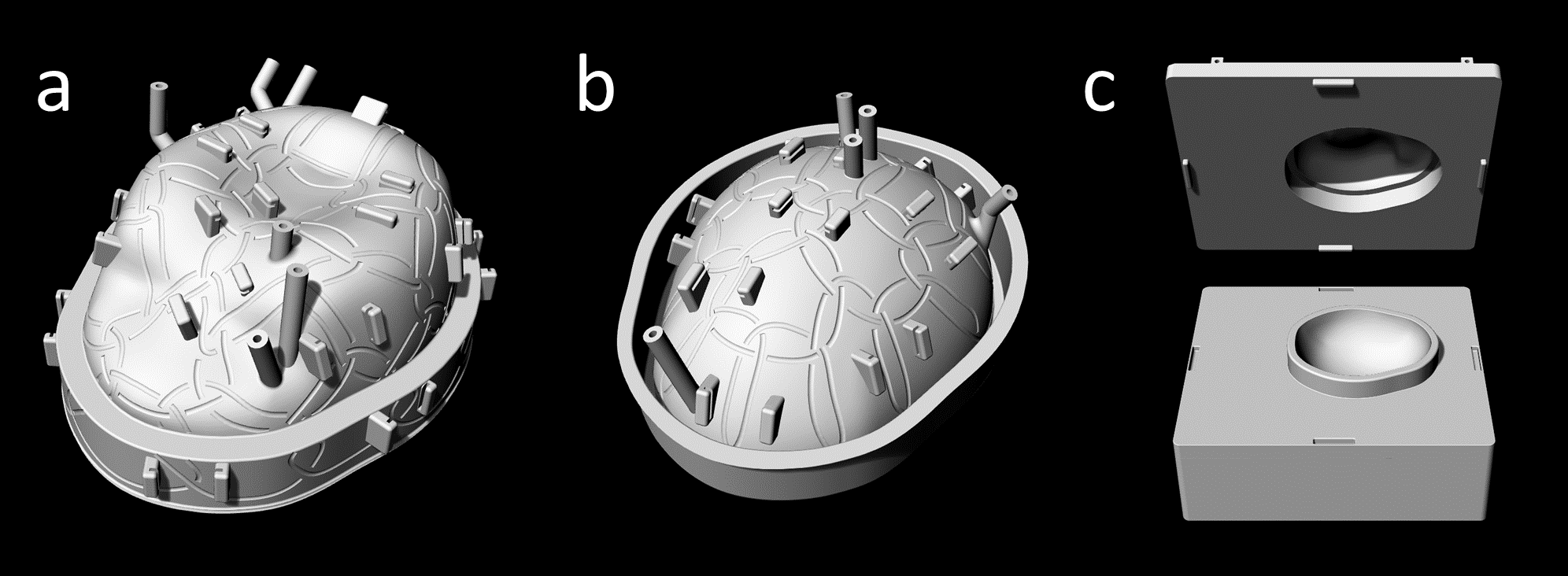
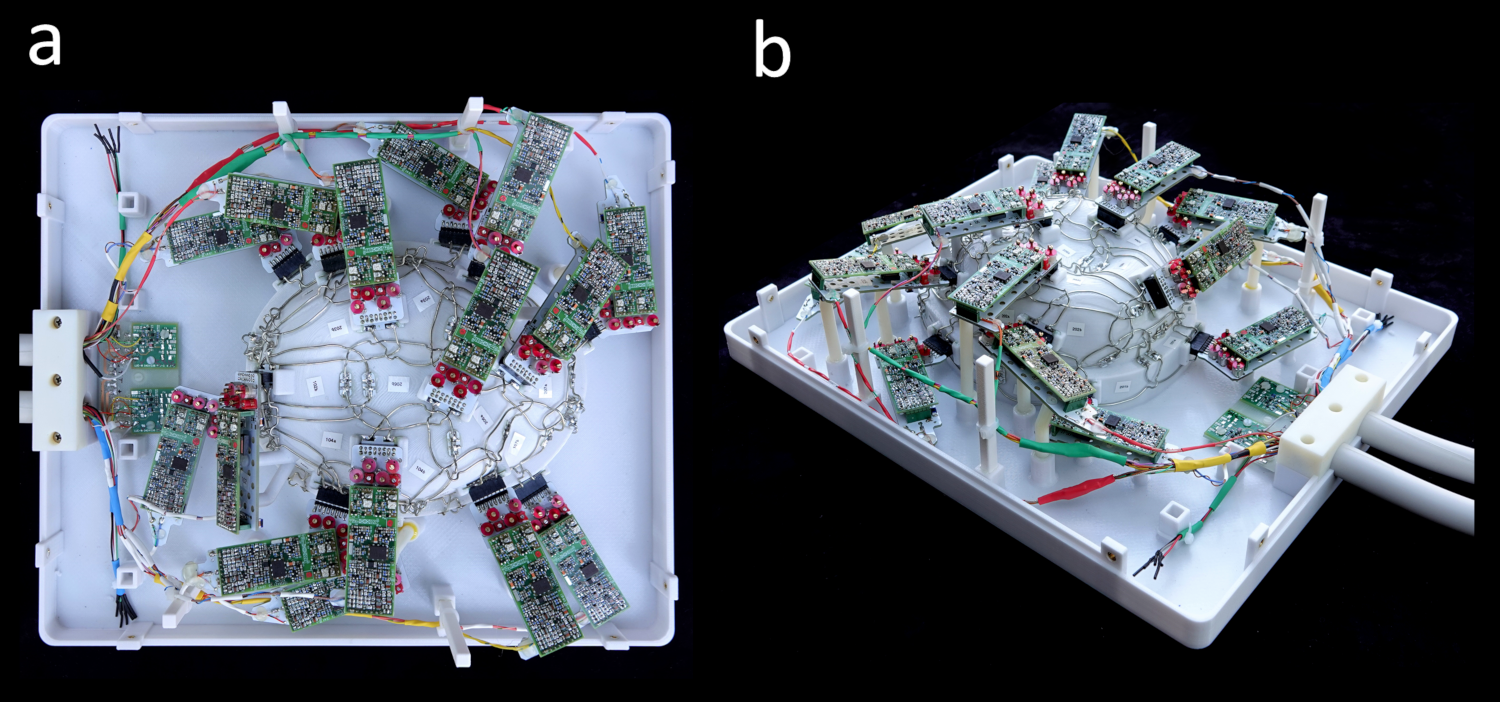
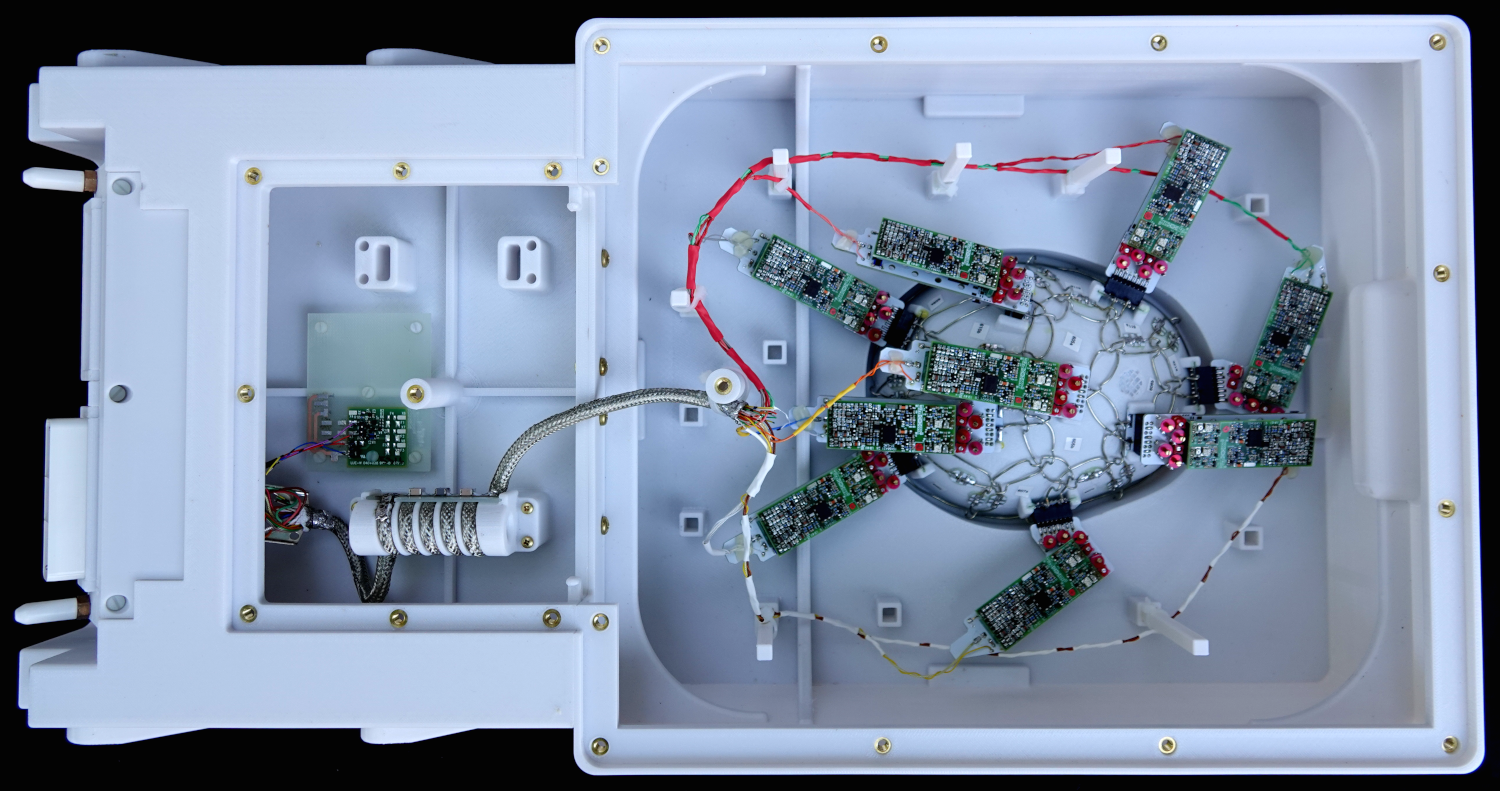
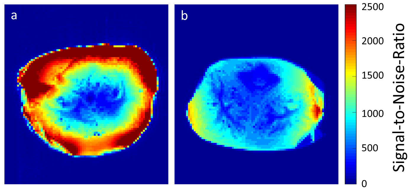
The optimized shape of the coilformer was derived from the modelled based on a non-linear atlas of the International Consortium for Brain Mapping (ICBM atlas). The layout of the overlapped coil elements was derived from a hexagonal/pentagonal tiling pattern. All loops were printed onto the coilformer together with standoffs to mount the preamplifiers. The average loop diameter was 54 mm. All helmet parts including its covers were printed in polycarbonate plastic using a rapid prototyping 3D printer (Fortus 360, Stratasys Ltd., Eden Prairie, MN, USA). The coil housing was split into two segments (Figure 1). It comprises 30 elements on the top half (see Figure 2) and 18 on the bottom half (see Figure 3), made of 16 AWG wire loops. Bench measurements verified the element tuning, active detuning, nearest-neighbor coupling and preamplifier decoupling for each coil element. Additionally, QU/QL-ratio was obtained with a coil element under test within the populated but detuned array assembly. As a load, we used a fixed tissue brain sample in the periodatelysine-paraformaldehyde (PLP) solution. Pairs of coils were attached to a twin preamplifier and low-noise converters (Siemens, Healthineers, Erlangen, Germany). Initial imaging was carried out on a 3T Connectome Scanner (Siemens Healthineers, Erlangen, Germany). SNR, noise correlation, and G-factor calculations were calculated from PD weighted raw-data images and compared to a custom-built 64ch whole-head coil [3] (Fig 4 and 5). Time course stability was derived from a EPI sequence (500 time points, TR/TE/FA= 1000 ms/30 ms/90°, M: 64 x 64, FOV: 200 x 200 mm2, SL: 5 mm, BW:2298 Hz/Pixel) [4].Results
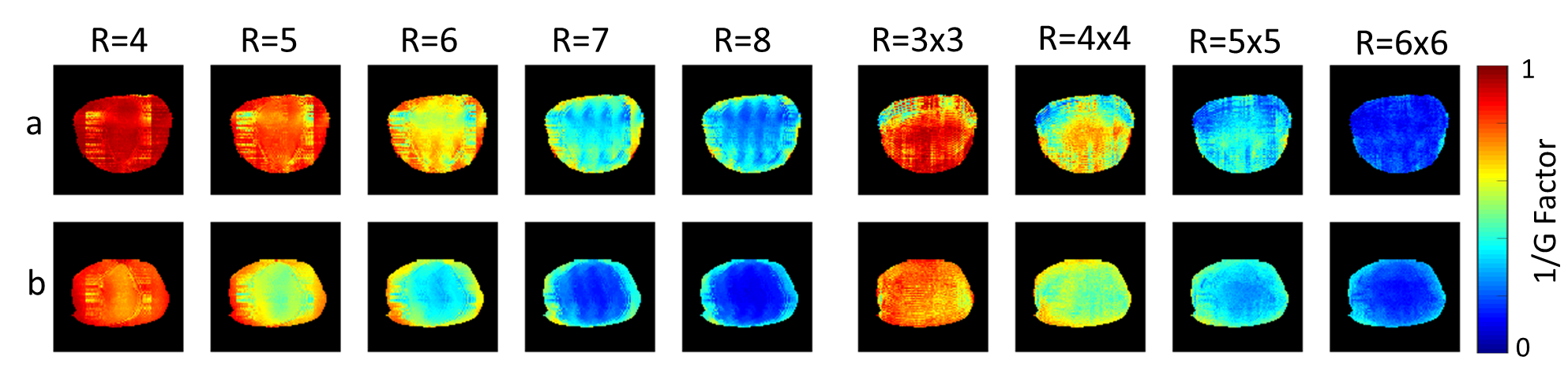
The coil elements showed a QU/QL-ratio of 233/46=5.1. Thus, the coil elements show sample noise domination. The average geometrical decoupling of nearest neighbors was measured to be 16 dB (12 dB to 21 dB). The coupling between non-adjacent pairs ranged from 11 dB to 28 dB. All coils were further isolated by 15 dB using preamplifier decoupling. Active detuning provided >42 dB isolation. The noise correlation between the channels ranged from 0.01% to 36% (avg. ~8%). Stability tests indicated a peak-to-peak variation of 0.4% over 500 time-points EPI sequence. The SNR gain (Figure 4) of the constructed 48ch coil in the cortical region was measured to be 2.1-fold, when compared to the 64ch in vivo head coil. The central SNR was equally observed for both coils. Figure 5 shows the inverse G-factor maps in transverse brain planes for 1D and 2D. The lower G-factors of the 48ch ex-vivo coil, roughly provides one additional unit of acceleration for a given noise amplification factor, when compared to the 64ch head coil.Discussion
Commonly used large channel count human head coil for ex-vivo whole brain sample studies renders brain imaging suboptimal. The customized 48ch coil, addresses this limitation by providing surrounding coil elements to the close proximity of the ex-vivo brain, thus providing increased reception sensitivity. The close-fitting and smaller elements of size-optimized offers a stronger spatial modulation of signal intensity and thus improved ability to unalias folded images (SENSE method) or synthesize spatial harmonics (GRAPPA methods), even when compared to a higher channel count head coil (slightly larger coil, no brain-shape optimized). Compared to literature [5], the unloaded-to-loaded Q-ratio shows increased sample noise fraction by a given loops size, when compared the coil elements optimized for in-vivo applications. This means, that the minimum diameter coil, which is sample noise dominated decreases in imaging fixed tissue brain samples in PLP solution; allowing us to contemplate very high-density arrays for ex-vivo sample imaging.Conclusion
A 48ch ex-vivo brain array coil was designed, constructed, and validated. The coil was characterized with both bench and image metrics and compared to a 64ch whole head coil. The coil is well-suited for high resolution ex-vivo MRI studies.Acknowledgements
Partially funded by BMBF grant # IN2016-2-226 and NIH grant # R01EB021265References
[1] Setsompop K, et. al. “Pushing the limits of in vivo diffusion MRI for the Human Connectome Project.” Neuroimage. 2013 Oct 15;80:220-33.
[2] McNab, JA, et al. “The Human Connectome Project and beyond: initial applications of 300 mT/m gradients.” Neuroimage 80 (2013): 234-245.
[3] Keil, B., et al. "A 64‐channel 3T array coil for accelerated brain MRI." Magnetic resonance in medicine 70.1 (2013): 248-258.
[4] Weisskoff, Robert M. "Simple measurement of scanner stability for functional NMR imaging of activation in the brain." Magnetic resonance in medicine 36.4 (1996): 643-645
[5] Keil, Boris, et al. "Size‐optimized 32‐channel brain arrays for 3 T pediatric imaging." Magnetic Resonance in Medicine 66.6 (2011): 1777-1787.
Figures