1485
Attachable B0-shim Array: An Add-on for RF Coils1Interdisciplinary Institute of Neuroscience and Technology, Qiushi Academy for Advanced Studies, College of Biomedical Engineering & Instrument Science, Zhejiang University, Hangzhou, China, 2MR Collaboration Northeast Asia, Siemens Healthcare, Hangzhou, China
Synopsis
In the present study, as an add-on to commercial-available RF coils, a novel B0-shim coil design for local B0 shimming was proposed, based on which 8-channel B0-shim array has been designed to attach to Nova 1Tx/32Rx head coil for local B0 shimming at 7T. With minor interference to RF coil, the apparent improvement in local B0 shimming has been demonstrated by using the proposed attachable B0-shim array. The present setup offers a feasible and promising means for practical higher-order local B0 shimming technology. It is believed that the proposed approach could potentially merit a broad scope of researches such as clinical diagnosis and cognitive neuroscience.
Introduction
Image distortion and signal dropout caused by B0 inhomogeneity has been a severe problem confronted in high-resolution functional imaging at ultra-high fields. Multi-coil B0-shim array has demonstrated its advantage in achieving higher order (>2) B0 shimming and mitigating local B0 inhomogeneity near nasal cavity and orbit [1-4]. However, for most multi-coil B0-shim arrays, their DC component was either integrated with the RF coils circuitry (restricted to the RF element), or physically independent to the RF coils – under both circumstances, RF coils have to be specially designed, which raises the complexity. In the present study, as an add-on to commercial-available RF coils, a novel B0-shim coil design for local B0 shimming was proposed, based on which 8-channel B0-shim array has been designed to attach to Nova 1Tx/32Rx head coil for local B0 shimming at 7T. The RF interference as well as the efficacy in improving local B0 homogeneity have been quantitatively evaluated over a physical phantom and a human subject.Method
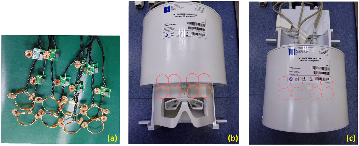
The 8-channel B0-shim array was constructed as eight loops with 3cm diameter and in two turns by using 21 AWG insulated cooper, as shown in Fig.1(a). Two toroid chokes were connected to each B0-shim loop to minimize coupled RF current. The toroid chokes were optimized to ensure the resonance frequency of individual B0-shim loop at least 70 MHz higher than the Larmor frequency (297.2MHz). Such an array was then attached to the surface of the inner helmet of Nova 1Tx/32Rx head coil (Nova Medical, MA, USA) on top of the forehead region, as indicated in Fig.1(b)(c). An 8-channel open-source, low-cost DC current amplifier board was used to generate independent shim current to each DC loop [5].
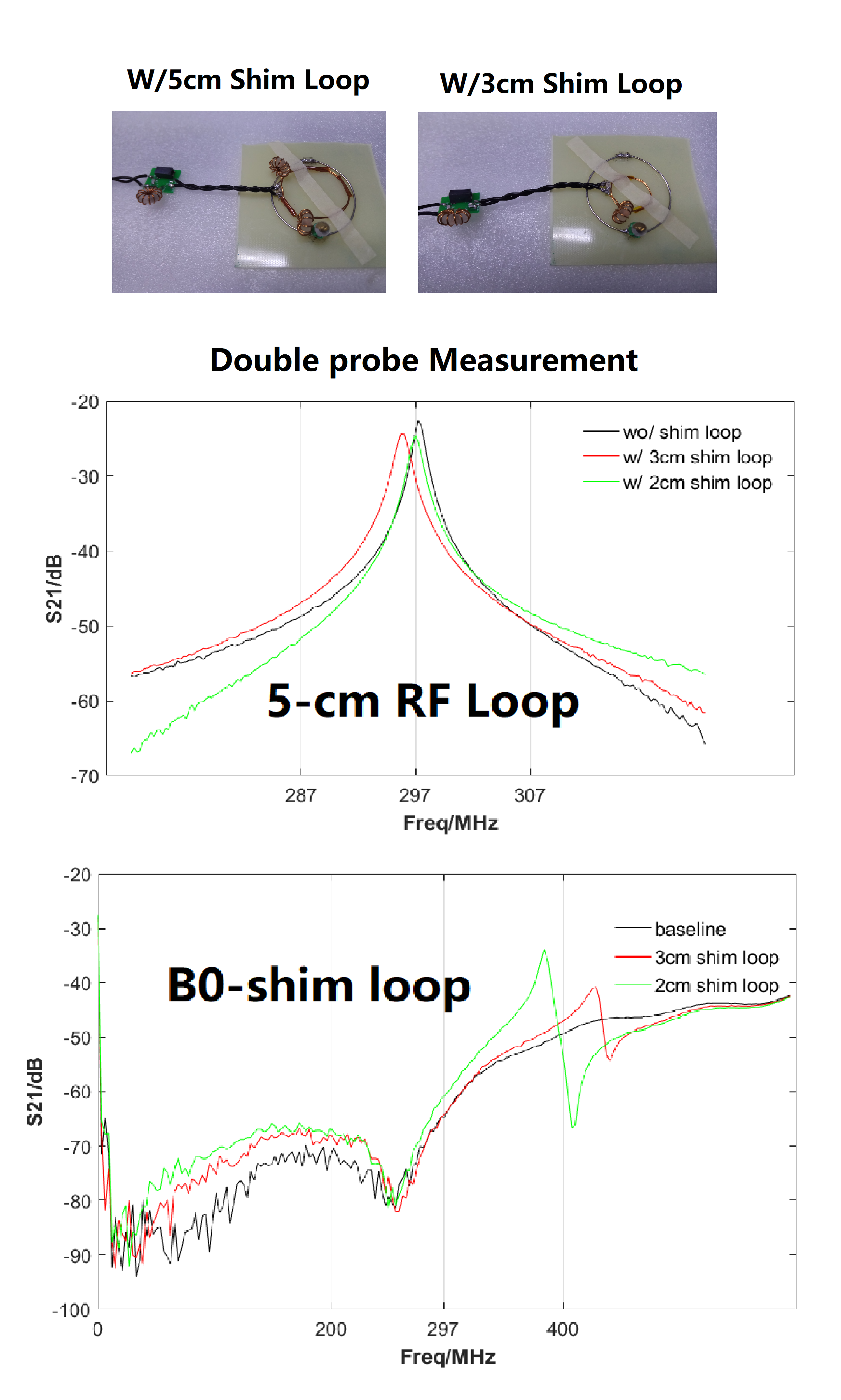
The RF interference introduced by the proposed B0-shim loop was evaluated on the bench through measuring S21 using the double probe and quality factor of an RF loop (5cm diameter) without vs. with B0-shim loops mounted, and S21 of B0-shim loops were also compared in two sizes: 3cm with 2 chokes vs. 2cm with 1 choke.
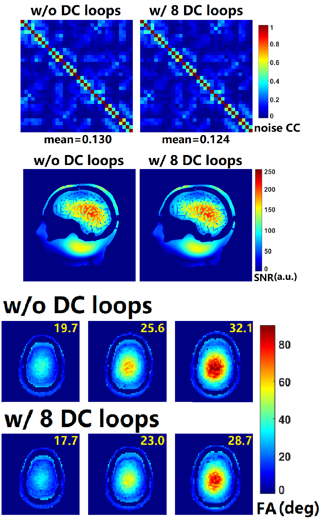
Images were acquired on a 7T research scanner (Magnetom, Siemens Healthcare, Erlangen, Germany) with a whole-body gradient set (70mT/m and 200T/m/s). A head-shape anthropomorphic Duke head phantom was used to collect coil Q&A measurements [6], and one healthy volunteer (30-year-old, 70 kg) was the subject to evaluate local B0 shimming efficacy (the protocol was approved by the local ethics committee, and a written consent was obtained from the subject.). System 2nd-order B0 shimming setting was applied prior to all scans. Before and after the B0-shim array was mounted, SNR and g-factor maps were obtained from PD-weighted FLASH images (TR/TE/α: 30ms/6ms/10°, 1×1×3mm3), and AFI-B1 maps [TR1/TR2/TE/α: 20ms/50ms/2.93ms/60°] were acquired to evaluate the transmission efficiency and homogeneity; the system reference voltage was set the same all the time.
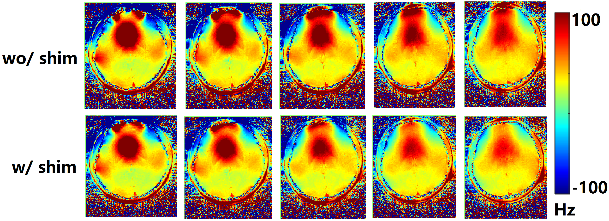
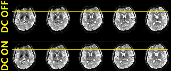
Furthermore, single-shot EPI images [TR/TE/α: 2,000ms/20ms/80°; matrix size 138×104; FOV 138×104mm2; slice thickness 2mm; bandwidth: 1,449Hz/px] with shim currents OFF/ON were acquired and compared. B0 field maps [TR/TE1/TE2/α: 429ms/3.29ms/4.31ms/65°; matrix size 138×104; FOV 138×104mm2; slice thickness 2mm; Distance factor: 100%] were also acquired with and without multi-coil B0 shim.
Results
Fig.2a showed that 0.3MHz and 1MHz frequency shift were introduced to the 5cm RF coil when 2cm and 3cm B0-shim loop were attached, respectively. The resonance frequency of both B0-shim loops was >80MHz higher than Larmor frequency, as in Fig.2b.
Fig.3 showed that less than 5% changes was introduced in the noise correlation matrix after adding the B0-shim array (row 1); besides, no apparent SNR change / deterioration was observed (row 2). On the transmit side, however, moderate decrease (~10%) in transmit efficiency was observed (row 3&4).
With multi-coil enabled B0 shimming capability, B0 shift around prefrontal lobe was significantly lowered as shown in Fig.4, and “notorious” signal dropout and image distortion within prefrontal lobe in single-shot EPI images have been effectively mitigated, as in Fig.5. Note that the DC current amplitude in each DC loop was constrained below 1A in our initial attempts.
Discussion and Conclusion
The apparent improvement in local B0 shimming has been demonstrated by using the proposed attachable B0-shim array. Minor RF interference may possibly be further mitigated by using smaller diameter loops and optimized chokes. The present setup offers a feasible and promising means for practical higher-order local B0 shimming technology; moreover, specialized designs can be adapted to various commercial-available RF coils at different field strengths. It is believed that the proposed approach could potentially merit a broad scope of researches such as clinical diagnosis and cognitive neuroscience.Acknowledgements
National Natural Science Foundation 81701774 and 61771423.References
[1] Juchem, Christoph, et al. "Dynamic multi-coil technique (DYNAMITE) shimming for echo-planar imaging of the human brain at 7 Tesla." Neuroimage 105 (2015): 462-472.
[2] Truong T K, Darnell D, Song A W. Integrated RF/shim coil array for parallel reception and localized B0 shimming in the human brain[J]. NeuroImage, 2014, 103: 235-240.
[3] Stockmann J P, Witzel T, Keil B, et al. A 32-channel combined RF and B0 shim array for 3T brain imaging. Magn Reson Med, 2015, 75(1): 441-451.
[4] Stockmann, Jason P., and Lawrence L. Wald. "In vivo B0 field shimming methods for MRI at 7 T." NeuroImage 168 (2018): 71-87.
[5] Arango, N., et al. "Open-source, low-cost, flexible, current feedback-controlled driver circuit for local B0 shim coils and other applications." Int Soc Magn Res Med. Vol. 1157. 2016. [6] http://phantoms.martinos.org/Main_Page
Figures