1478
Passive shimming for a Portable Head-Only scanner1Robinson Research Institute, Faculty of Engineering, Victoria University of Wellington, Lower Hutt, New Zealand, 2Department of Biomedical Engineering, Columbia University in the City of New York, New York, NY, United States, 3Center for Magnetic Resonance Research, Department of Radiology, University of Minnesota, Minneapolis, MN, United States, 4Department of Radiology, Columbia University in the City of New York, New York, NY, United States
Synopsis
Part of a collaboration under NIH grant U01EB025153 is leading to the development of a highly compact brain imaging scanner. In order to realize the required B0 uniformity of the scanner, we have developed passive shimming techniques that will be robust despite a low field uniformity and non-cylindrical magnet warm bore layout. This presentation describes the methods and results we will use to shim our compact scanner.
Introduction
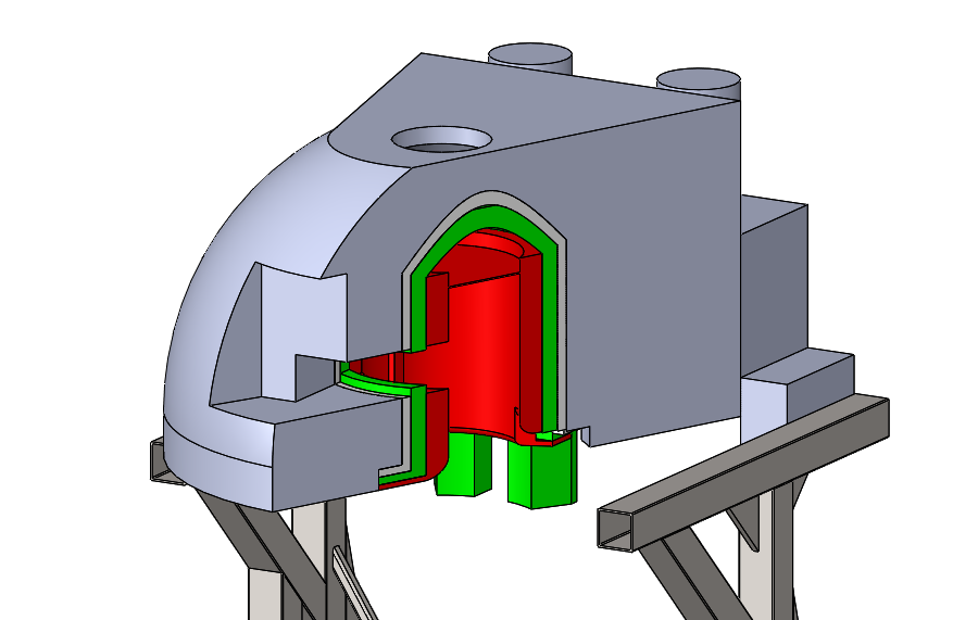
Using novel MRI methods1,2,3 embedded in the U01-EB025153 collaboration, we have demonstrated capability to generate MRI images in poor B0 uniformity. Victoria University of Wellington has subsequently designed a compact 1.5 T magnet, shown as part of a system in Figure 1. The magnet has ±10kHz B0 uniformity over an elliptical region of interest (ROI) sized 15x20x15 cm. The ROI is designed to encompass an average human brain with B0 uniformity compatible with our imaging techniques. The reduced uniformity and tapered warm bore has allowed the magnet to be designed in a very compact way, completely eliminating the shoulders from within the magnet. The design also includes a patient window, enabling natural communication with the subject during image acquisition.
As is well known, upon manufacture of an MRI magnet, field uniformity is substantially degraded compared to the designed uniformity. Passive shimming has been routinely used in clinical scanners to correct the difference in uniformity between the design and as-manufactured uniformity4. Our magnet solution’s unique features are enabling technologies for advanced motor coordination studies, but when coupled to a non-uniform magnetic field represent a new set of challenges for existing passive shimming methods. Here we extend conventional passive shimming practice to consider when passive shims must be limited by magnet geometry to a non symmetric distribution.
Methods
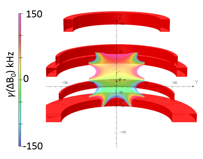
From an earlier design iteration of the magnet shown in Figure 1, where the field uniformity was closer to ± 100 kHz over the ROI, we designed and built a low temperature superconductor magnet at half scale to the final magnet design. The magnet design and design uniformity is shown in Figure 2.
We have previously developed and demonstrated passive shim software in MATLAB5 to passively shim magnets with a conventional cylindrical bore geometry. We modified this software to allow an arbitrary shim geometry.
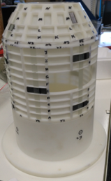
We designed and 3D printed the cassette shown in Figure 3 from SLA material to accommodate the shim pieces. The cassette included the same taper as in the magnet shown in Figure 1 and has a window aligned with the window through the magnet, albeit at half scale.
The B0 field from the magnet was measured in Tesla over the surface of a 120 mm diameter sphere using a Hall effect sensor moved by a linear actuator. For measurement purposes, the sphere was decomposed into a series of 12 axial planes on which a series of 10 equally radially spaced measurements were taken at the intersection of the sphere and the axial planes.
Spherical harmonic terms were then fitted to the B0 measurements and an optimal passive shim configuration was computed compatible with the pocket sizes and locations on the passive shim cassette.
Our initial design approach was to load a set of ferromagnetic shims to set all spherical harmonic terms less than 3rd order to be zero.
Results
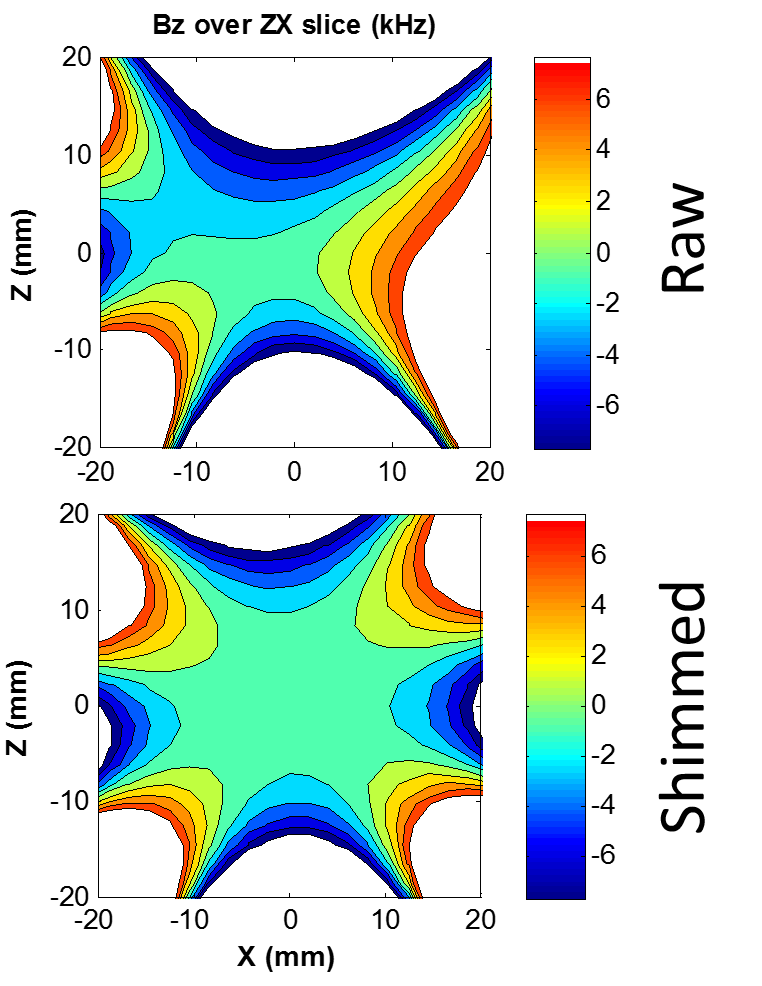
The initial field uniformity as measured by the Hall effect sensor is shown in the top half of Figure 4. For clarity, a limited section of the 7.5x10x7.5 cm elliptical ROI has been shown. By convention, the z axis is the solenoidal axis of the magnet and the +y direction is the axis along which the window is located.
We calculated where the ferromagnetic shims needed to be loaded, loaded the shims and re-measured the field. Upon repeating the measurement and passive shim loading process five times, the spherical harmonic terms of B0 had converged to their design values. Strictly, the shimming process had converged after the first three iterations, with the remaining iterations confirming the process convergence.
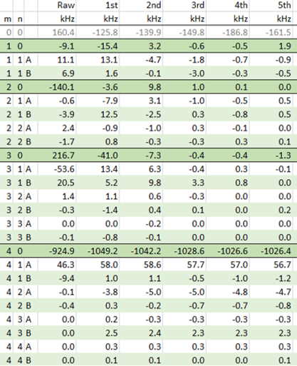
The field uniformity was improved as can be seen in the lower half of Figure 4. The improvement in uniformity in terms of individual spherical harmonics terms is summarized in Figure 5. The level of improvement is consistent with the number of spherical harmonic orders that were targeted; as expected, higher order B0 non-uniformity now dominates.
Conclusion and Outlook
We have developed a passive shimming approach that allows us to improve the field uniformity of a half-scale replica of a soon-to-be built compact head-only scanner. The shimming approach converges on a target field despite the presence of a patient window and taper along the solenoidal direction of the magnet, providing predictable shimming outcomes.
The extension of this method to higher than second order terms is straightforward and expected to provide further improved field uniformity; however, the next stage of our work is to develop passive shimming methods that target an ellipsoidal rather than spherical imaging volume, and that will allow peak-to-peak shimming methods in addition to harmonic approaches.
Acknowledgements
This research was supported by the National Institute of Biomedical Imaging and Bioengineering of the National Institutes of Health under award number U01EB025153.References
1. Patz S, Wong STS, Roos MS. Missing pulse steady‐state free precession. Magn Reson Med. 1989;10(2):194-209.
2. Kobayashi N, Idiyatullin D, Adriany G, Garwood M. Magnetic Resonance Imaging under Highly Inhomogeneous B0 Fields using Missing-Pulse Steady-State Free Precession (MP-SSFP). In: Proc. Intl. Soc. Magn. Reson. Med. 25, 5052 (2017).
3. Snyder ALS, Corum CA, Moeller S, Powell NJ, Garwood M. MRI by steering resonance through space. Magn Reson Med. 2014;72(1):49-58.
4. Belov A, Bushuev V, Emelianov M, Eregin V, Severgin Y, Sytchevski S and Vasiliev V. Passive shimming of the superconducting magnet for MRI IEEE Trans. Appl. Supercond. 1995; 5 679–81
5. Parkinson B J, Slade R A, Bouloukakis K. A compact 3 T all HTS cryogen-free MRI system. Supercond. Sci. Technol. 2017. 30 125009
Figures