1472
High-resolution numerical simulation of respiration-induced dynamic B0 shift in the head in high-field MRI1Department of Biomedical Engineering, Sungkyunkwan university, Suwon, Korea, Republic of, 2Center for Neuroscience Imaging Research, IBS, Suwon, Korea, Republic of
Synopsis
To investigate B0 fluctuation in the head induced by respiration in high field MRI, we simulated respiration with a human 4D phantom model, and calculated B0 in the brain by an efficient calculation algorithm. Simulated B0 was analyzed for the spatiotemporal distribution and voxel size dependence. The amplitude of dynamic B0 change exhibited strong inferior/superior gradient and significant anterior/posterior gradient, consistent with previous experimental data. Compared to the previous modeling studies, our simulation can yield more reliable, high-resolution results within a relatively short calculation time.
Introduction
In high-field MRI, B0 shift induced by tissue susceptibility is a significant source of imaging artifacts. In particular, respiration-induced B0 shift in the head has been much studied due to its importance in functional MRI at ultra-high magnetic fields (1,2). Previous studies measured B0 shift using gradient-echo phase imaging (3), magnetic field probes (4), or from post-processing of multi-channel images (5). These works had a few limitations, such as low (temporal or spatial) resolutions and a small number of subjects, with a limited range of anatomies and breathing conditions. In this work, we propose high-resolution simulation of respiration-induced B0 shift in the head using a detailed 4D human body model, and an artifact-free B0 calculation algorithm gSVC (generalized susceptibility voxel convolution) (6). We obtained B0 shift in the head with 1 to 10 mm isotropic voxel sizes through a single respiration cycle, and analyzed the spatiotemporal B0 variation and voxel size dependence.Methods
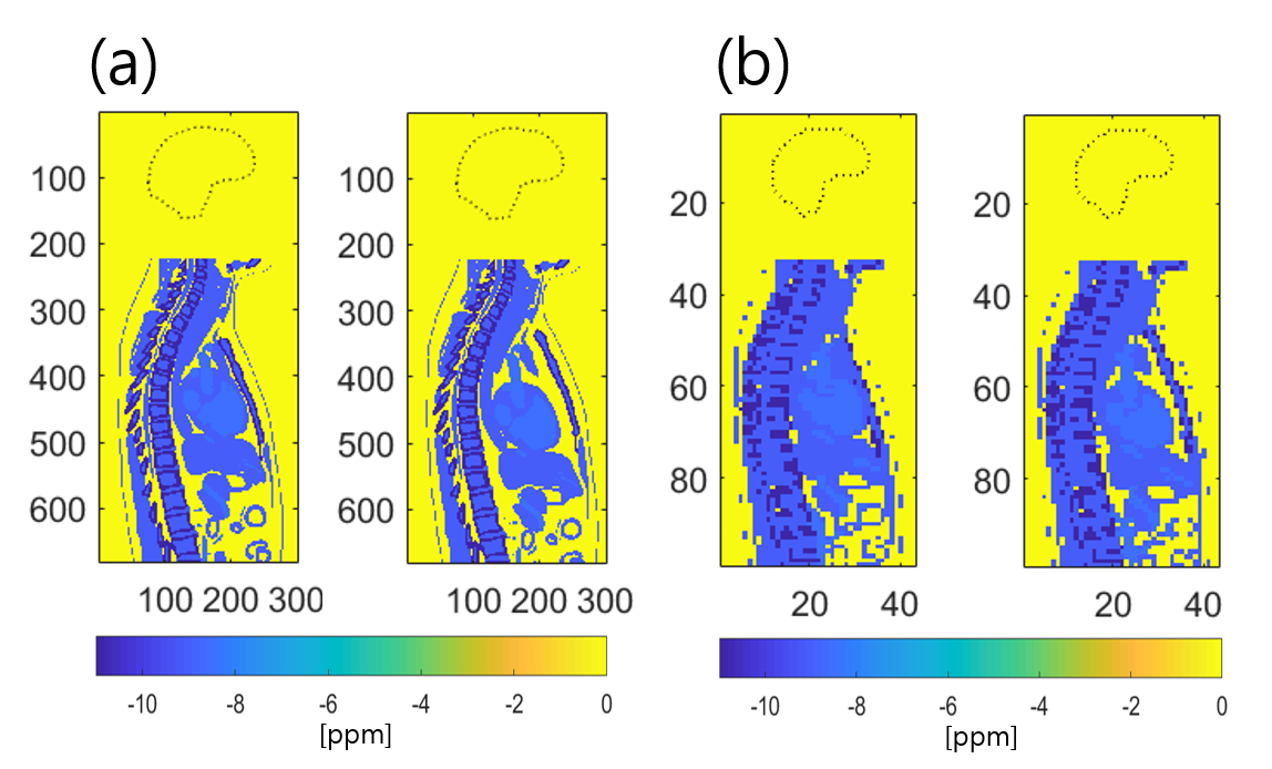
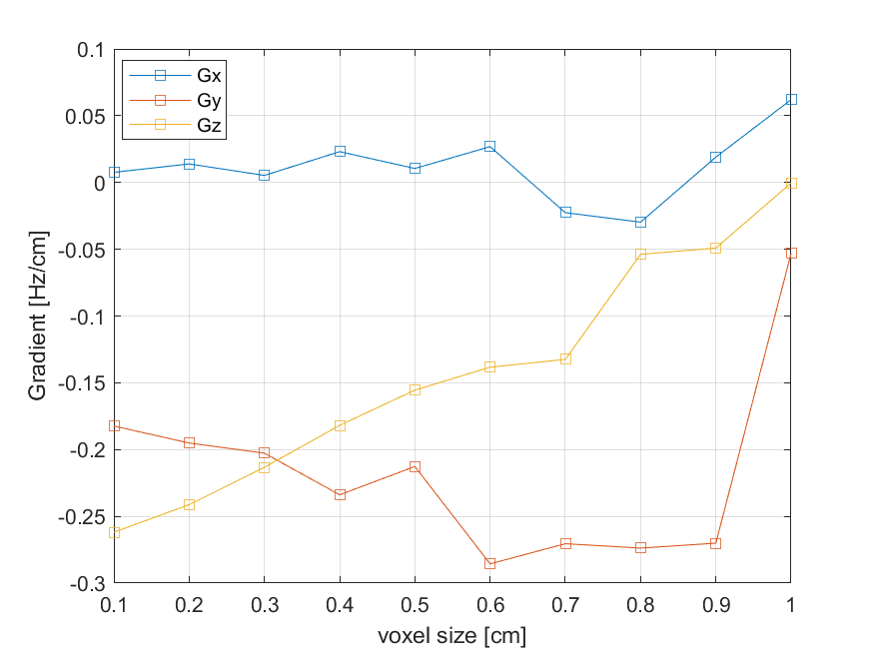
The 4D human body model was taken from XCAT (extended cardiac-torso model) (7-11), which allows adjusting anatomical and dynamic motion parameters. In this work, we used the following parameters: gender = male, voxel size = 1 to 10 mm (isotropic), body length = 560 mm (from L2 to C3, susceptibility = 0 (air and lung)/ -8.5 (blood) / -11 (bone) / -9 (other tissues) ppm, maximum diaphragm motion = 2.0 cm, maximum anterior/posterior expansion = 1.2 cm (normal breathing). The respiration cycle was 5 seconds, starting from the exhaled position with a total of 10 frames. The main magnetic field was set to be 7 T. The gSVC method was used instead of more conventional k-space-discretized B0 calculation algorithm for computational efficiency and artifact reduction. All calculations were performed in Matlab (R2017b, Mathworks, USA) on a 64 bit Windows PC with 64 GB RAM and Intel Xeon(R) CPU E5-1607 v4. The computational times for the dipolar field kernel calculation (t1) and dynamic B0 update (t2) were recorded for each voxel size. The B0 map of the first frame was subtracted from the subsequent frames, and the resulting B0 shift maps were analyzed in terms of their gradients: Gx = dB0/dx, Gy = dB0/dy, Gz = dB0/dz, where x, y, z are coordinates in the left to right, posterior to anterior, and the feet to head directions, respectively.Results
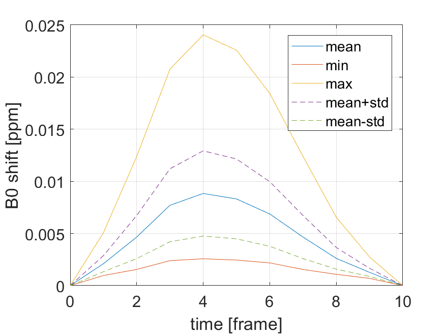
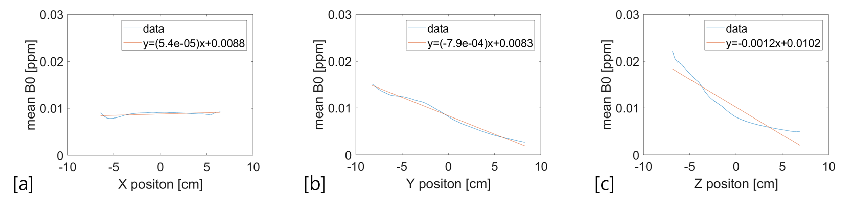
Figure 1 shows mid-sagittal slices of fully exhaled and inhaled positions in the simulation. Figure 2 shows that the maximum B0 change is about 0.024 ppm (7.2 Hz at 7T) at the time of full inhalation. This is comparable to a recent experimental data (5). Over the whole brain, the calculated mean B0 shift at the peak variation was about 0.008 ppm (2.4 Hz). The sign of the change is consistent with the lung filled with air, which is more paramagnetic than the tissue. Following the analysis of ref (3), the B0 values on each slice normal to each of the gradient directions (x, y, z) were averaged at the fully inhaled position. The 2D averaged B0 was fitted to straight lines and plotted against the coordinates in the gradient directions (Fig.3). The magnitude of B0 change declined as one moved in the superior and the anterior directions. This trend agrees with the previous experimental results (3). Figure 4 shows that in our model, voxel sizes greater than 5 mm are unreliable as B0 gradient values become more erratic. Table 1 shows that the total computation time (t1 + t2) for 1 mm isotropic voxel was only 90 seconds. In our model, 2 mm voxel size might represent a good balance between the simulation accuracy and computational time.Discussion
Compared to the previous simulation work (12), we implemented up to 7.63 = 439 times smaller voxel volume, thereby reflecting more details of organ movements associated with respiration. The human body model used allows variation of many anatomical and physiological parameters corresponding to different breathing conditions, which may be of use in future studies on dynamic shimming. In conclusion, we have shown that respiration-induced B0 shift in the head can be computed with high spatiotemporal resolution by utilizing a detailed dynamic human body model with an efficient B0 calculation algorithm. Future work includes verifying our results with 4D susceptibility models derived from actual human lung images (13), and developing methods to compensate the respiration-induced B0 shift by dynamically controlled coils or magnetic materials.Acknowledgements
This work was supported by IBS-R015-D1.References
1. Zeller M, Kraus P, Muller A, Bley TA, Kostler H. Respiration impacts phase difference-based field maps in echo planar imaging. Magn Reson Med 2014;72(2):446-451.
2. Zahneisen B, Asslander J, LeVan P, Hugger T, Reisert M, Ernst T, Hennig J. Quantification and correction of respiration induced dynamic field map changes in fMRI using 3D single shot techniques. Magn Reson Med 2014;71(3):1093-1102.
3. Van de Moortele PF, Pfeuffer J, Glover GH, Ugurbil K, Hu X. Respiration-induced B0 fluctuations and their spatial distribution in the human brain at 7 Tesla. Magn Reson Med 2002;47(5):888-895.
4. Vannesjo SJ, Wilm BJ, Duerst Y, Gross S, Brunner DO, Dietrich BE, Schmid T, Barmet C, Pruessmann KP. Retrospective correction of physiological field fluctuations in high-field brain MRI using concurrent field monitoring. Magn Reson Med 2015;73(5):1833-1843.
5. Meineke J, Nielsen T. Data-driven correction of B0-off-resonance fluctuations in gradient-echo MRI. 26th Annual Meeting of ISMRM. Paris, France 2018. p 1172.
6. Lee SK, Hwang SH, Barg JS, Yeo SJ. Rapid, theoretically artifact-free calculation of static magnetic field induced by voxelated susceptibility distribution in an arbitrary volume of interest. Magn Reson Med 2018;80(5):2109-2021.
7. Segars WP, Mahesh M, Beck TJ, Frey EC, Tsui BM. Realistic CT simulation using the 4D XCAT phantom. Med Phys 2008;35(8):3800-3808.
8. Silva-Rodriguez J, Tsoumpas C, Dominguez-Prado I, Pardo-Montero J, Ruibal A, Aguiar P. Impact and correction of the bladder uptake on 18 F-FCH PET quantification: a simulation study using the XCAT2 phantom. Phys Med Biol 2016;61(2):758-773.
9. Koybasi O, Mishra P, St James S, Lewis JH, Seco J. Simulation of dosimetric consequences of 4D-CT-based motion margin estimation for proton radiotherapy using patient tumor motion data. Phys Med Biol 2014;59(4):853-867.
10. Lowther N, Ipsen S, Marsh S, Blanck O, Keall P. Investigation of the XCAT phantom as a validation tool in cardiac MRI tracking algorithms. Phys Medica 2018;45:44-51.
11. Paganelli C, Summers P, Gianoli C, Bellomi M, Baroni G, Riboldi M. A tool for validating MRI-guided strategies: a digital breathing CT/MRI phantom of the abdominal site. Med Biol Eng Comput 2017;55(11):2001-2014.
12. Marques JP, Bowtell R. Application of a fourier-based method for rapid calculation of field inhomogeneity due to spatial variation of magnetic susceptibility. Concept Magn Reson B 2005;25B(1):65-78.
13. Park J, Shin T, Yoon SH, Goo JM, Park JY. A radial sampling strategy for uniform k‐space coverage with retrospective respiratory gating in 3D ultrashort‐echo‐time lung imaging. NMR in biomedicine 2016;29(5):576-587.
Figures