1461
An adjustable susceptibility phantom for evaluation of field mapping and fat fraction estimation algorithms1Robarts Research Institute, the University of Western Ontario, London, ON, Canada
Synopsis
We demonstrate a method to produce phantoms with arbitrary and easily adjustable susceptibility distributions. Objects in a magnet bore cause high order inhomogeneities not correctable by shimming, and confound magnetic field mapping and phase unwrapping algorithms. We show a wire winding carrying direct current can simulate an object of adjustable susceptibility, for rapid testing of field mapping and phase unwrapping methods.
Introduction
Local B0 magnetic field homogeneity is critical in MRI but is difficult to achieve, particularly at high field strength, at interfaces, and in the presence of implanted objects. While higher order shims can compensate for much of the inhomogeneity, the ability to measure and compensate for B0 inhomogeneities remains of interest.1,2 Different algorithms for measuring B0, all relying on measurement of the phase, exist but are rarely compared against each other, in part, due to the difficulty of generating controlled inhomogeneity conditions. To test the performance of phase unwrapping and field mapping methods in the presence of high order field distortions we have developed and tested a method to produce arbitrary, variable, and controlled field distortions within the magnet bore, thereby simulating the insertion of objects of differing susceptibilities. Quantifying algorithm performance against known inhomogeneity will inform models of the effects of inhomogeneities, with the ultimate goal of enabling more effective correction of these effects.Methods
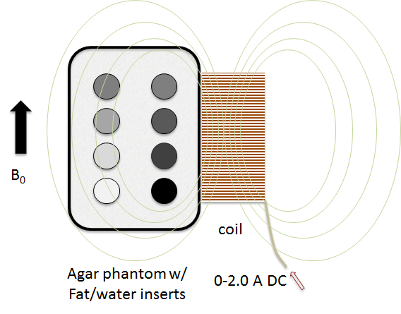
Variable susceptibility coil: A variable susceptibility object was simulated by winding 90 turns of 0.64 mm diameter insulated copper onto a 75 mm diameter, 100 mm cylindrical form. The resulting short solenoid can be used to approximate varying susceptibility by providing stable direct current through it when it is placed in the magnet bore, with the solenoid’s axis parallel to B0. A well-filtered stabilized DC source, placed outside the magnet room, was used to feed the solenoid through a low-pass line filter, a penetration-panel RF filter, and twisted-pair wiring. The winding current was varied between zero and 2 amperes, producing a field shift within the solenoid of up to 2 mT, resulting in an apparent susceptibility of 667 ppm at 3T.
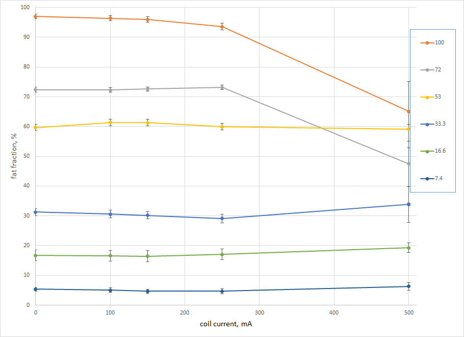
Test phantom: Following the method of Hattori et al.,3 a series of fat-water emulsions consisting of peanut oil and water, emulsified with SDS and Pluronics P123 were stabilized in 2% w/w agar, reducing T2 to approximately 60 ms. The emulsions were doped with GdCl3 to yield a T1 of approximately 1000 ms. Fat fractions of 100, 72, 53, 33, 17, 7.4, 2.5 and 0 percent were prepared and sealed in 50 mL centrifuge tubes, and then suspended in a 2-litre fat-free outer volume of 2% agar in a sealed plastic container. The susceptibility-simulating winding was taped to the exterior surface of the phantom, as shown in Fig. 1.
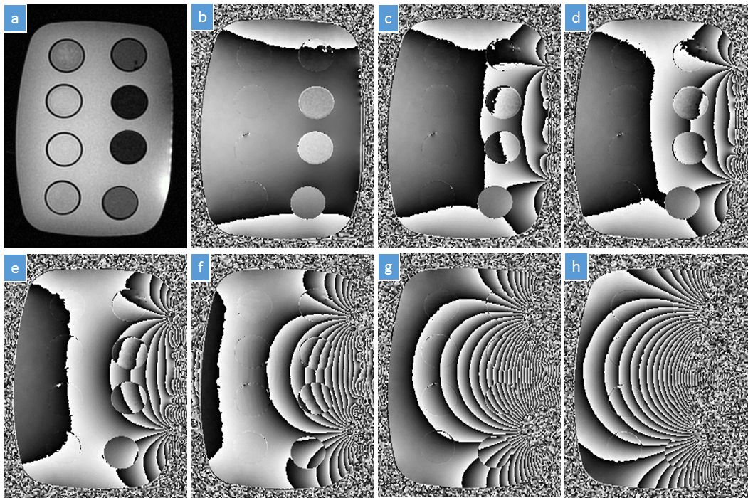
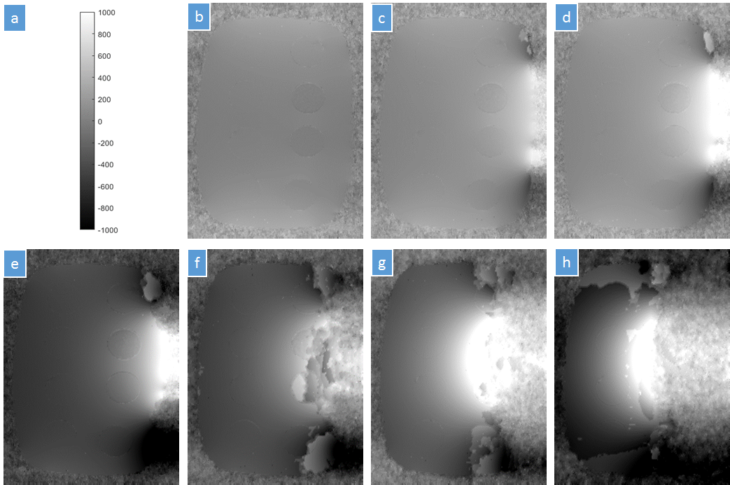
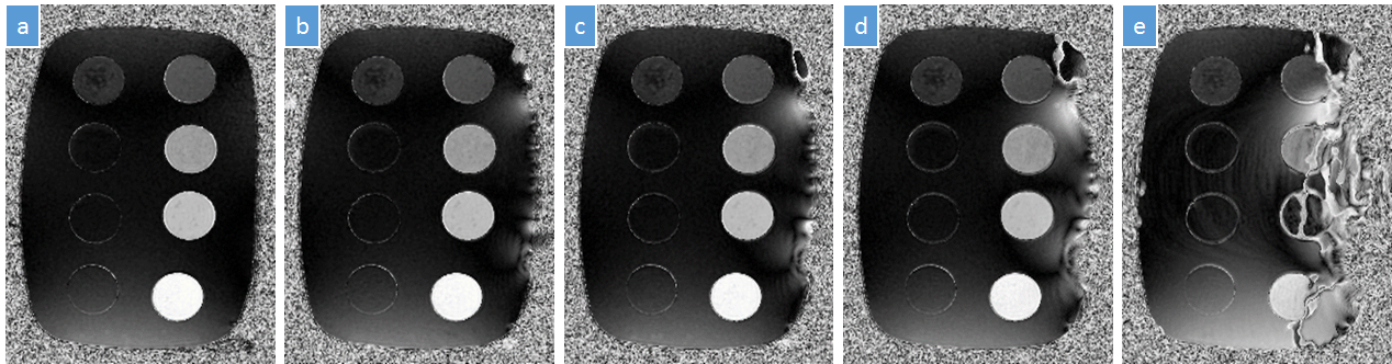
Imaging: MR imaging was performed on a clinical 3T whole-body scanner, using a 32-channel cardiac receive coil. A dual echo-train 3D GRE sequence (TE1/∆TE/TE5 = 3.20/1.46/9.04 ms, TE6/∆TE′/TE10 = 16.75/7.15/45.35 ms, TR: 47.6 ms, resolution: 1.04x0.78x3 mm3, matrix: 192x256x18, BW: 142.86 kHz, flip angle: 10°, scan time = 2 min 46 sec) optimized for fat fraction and R2* measurements was used to test the concept.4 Imaging was performed with coil current of 0, 100, 150, 250, 500, 1000 and 2000 mA. Data were reconstructed and processed offline. Phase unwrapping was performed using the PUROR algorithm5 and B0-NICE6 was used to generate a B0 map and a fat fraction map.
Results & Discussion
The field perturbation from the solenoid winding, evidenced by the phase maps of Fig. 2, appears qualitatively as expected, and is of sufficient magnitude to simulate materials of relatively large susceptibility. The field shift outside the solenoid in the vicinity of the phantom varies up to approximately 0.3 mT, producing a local gradient variable up to approximately 10-100 Hz/pixel depending on proximity to the winding (Fig. 3). The results of Figures 4 and 5 show the measured fat fractions at varying currents.
No interaction between the inserted winding and the magnet gradients was observed. A small amount of B1 RF field distortion may be present (not shown here); further work is needed to quantify its effect. Residual susceptibility of the coil materials appears to introduce small artifacts in the immediate proximity to the winding. Future designs may use low-susceptibility conductor and form materials to minimize the effect.
For this proof of concept, a simple solenoid was chosen for ease of construction, acknowledging that the apparent susceptibility of the simulated cylinder will not be uniform through the cylinder volume. However, in general, wire-winding patterns can be designed to simulate, with some limitations, arbitrary object shapes and susceptibility distributions.
Conclusion
We have demonstrated it is practical to produce a simple shape with adjustable apparent susceptibility in a magnet. We expect to use this approach to test methods to compensate for local susceptibility differences in a magnet bore.Acknowledgements
No acknowledgement found.References
1. Balachandrasekaran A, Mani M, Jacob M. Calibration-free B0 correction of EPI data using structured low rank matrix recovery. IEEE Trans Med Imaging 2018.
2. Ngo GC, Bilgic B, Gagoski BA, Sutton BP. Correction of magnetic field inhomogeneity effects for fast quantitative susceptibility mapping. Magn Reson Med 2018.
3. Hattori K, Ikemoto Y, Takao W, et al. Development of MRI phantom equivalent to human tissues for 3.0-T MRI. Med Phys 2013;40:032303.
4. Liu J, Christiansen SD, Drangova M. Single multi-echo GRE acquisition with short and long echo spacing for simultaneous quantitative mapping of fat fraction, B0 inhomogeneity, and susceptibility. Neuroimage 2018;172:703-717.
5. Liu J, Drangova M. Intervention-based multidimensional phase unwrapping using recursive orthogonal referring. Magn Reson Med 2012;68:1303-1316.
6. Liu J, Drangova M. Method for B0 off-resonance mapping by non-iterative correction of phase-errors (B0-NICE). Magn Reson Med 2015;74:1177-1188.
Figures