1459
Numerical assessment of a multi-coil shimming system in human brain MRI1Institute of Biomedical Engineering, National Taiwan University, Taipei, Taiwan, 2Taipei Medical University - Shuang Ho Hospital, Taipei, Taiwan, 3Department of Medical Biophysics, University of Toronto, Toronto, ON, Canada, 4Department of Neuroscience and Biomedical Engineering, Aalto University, Espoo, Finland
Synopsis
We numerically evaluated the performance of multi-coil shimming in human brain using an array of up to 1,000 coils with realistic off-resonance distributions of 37 healthy participants. The average and variation of shim current distributions were revealed. Singular Value Decomposition suggested orthogonal current modes to reduce off-resonance. The first 6 current modes accounted for about 90% of the variance of shim current distributions. They achieved shimming performance comparable to the 5th-order and 4th-order spherical harmonic in global and slice-selective shimming, respectively.
Introduction
To mitigate off-resonance artifacts, shim fields are created to reduce field inhomogeneity. The shim fields can be modeled by spherical harmonic (SH) functions1,2 or a collection of localized shim coils in multi-coil (MC) shimming3-5. Here we numerically evaluated the performance of MC shimming on the human brain using a shim coil array with up to 1,000 coils to reduce the off resonance in a group of participants (N=37). Specifically, we I) studied average and variation of shim current distributions, II) revealed important orthogonal “modes” of shim current distributions, III) evaluated how much off-resonance in human head can be reduced by different modes of shim current distributions, and IV) compared multi-coil shimming with SH shimming.Methods
The off-resonance field maps were measured on a 3T MRI (Skyra; Siemens) using a two-echo sequence (TE1 = 2 ms, TE2 = 4.46 ms) from 37 participants, who gave written informed consents approved by the Institution Review Board of National Taiwan University Hospital. Before the measurement, the MRI was first shimmed by the 2nd-order systematic shimming. Off-resonance field maps were co-registered to the standard brain MNI 305 atlas6. Shimming was simulated using an array consisting of circular shim coils evenly distributed over either a complete sphere (Ns = 1,000, 500, 200 or 100) or a partial sphere (Ns = 891, 452, 174 or 88). Figure 1 shows the shim arrays. The partial sphere removed coils in the bottom in order to create a circular space to accommodate the neck. The shim field generated by one circular shim coil was calculated by the Biot-Savart’s law. The off-resonance field from a participant was shimmed by a given shim array using ridge regression7,8 to avoid over fitting. We pooled 36 estimated shim current distributions from 36 participantsto create a matrix,which was subsequently decomposed by Singular Value Decomposition (SVD) into orthogonal “current modes”. The shimming performance of current modes from pooled data was evaluated by calculating the root-mean-squares of the residual field after using these current modes to shim the off-resonance field of the left-out participant. The data pooling and shimming was repeated 37 times across different left-out participants. We also evaluated the performance of slice-selective shimming. The shim fields at a specified axial slice was separately estimated based on the shim current pattern optimized for global shimming. For comparison, slice-selective shimming was also calculated using the 0th to the 7th-order of spherical harmonics9.Results
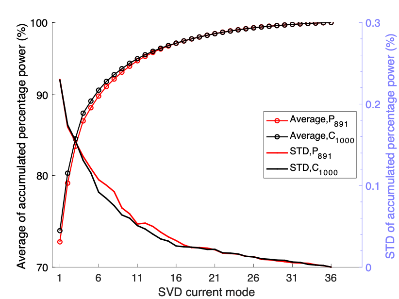
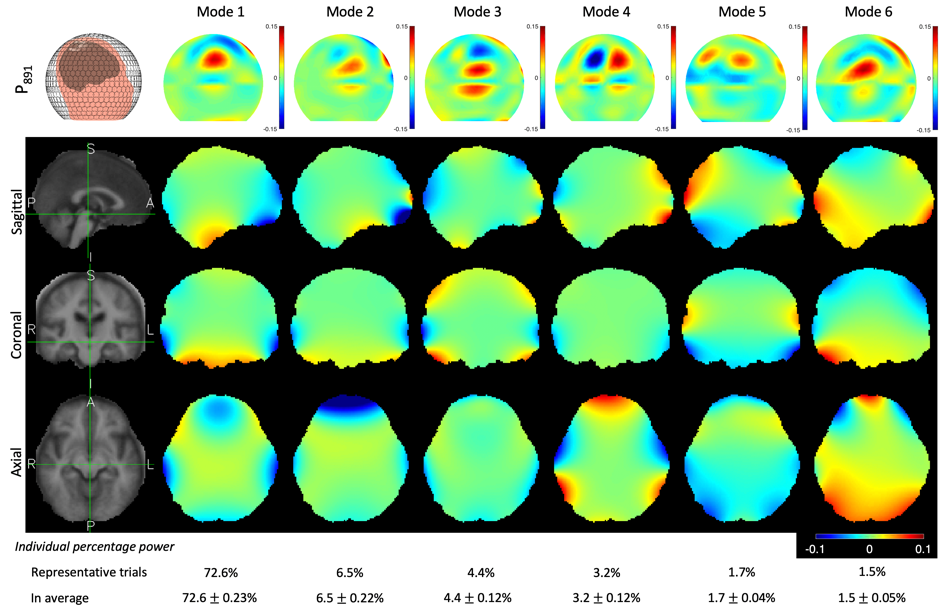
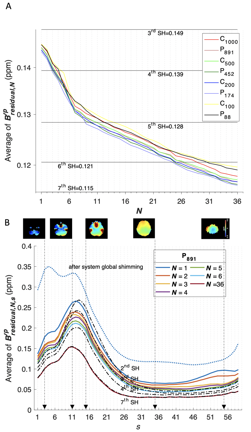
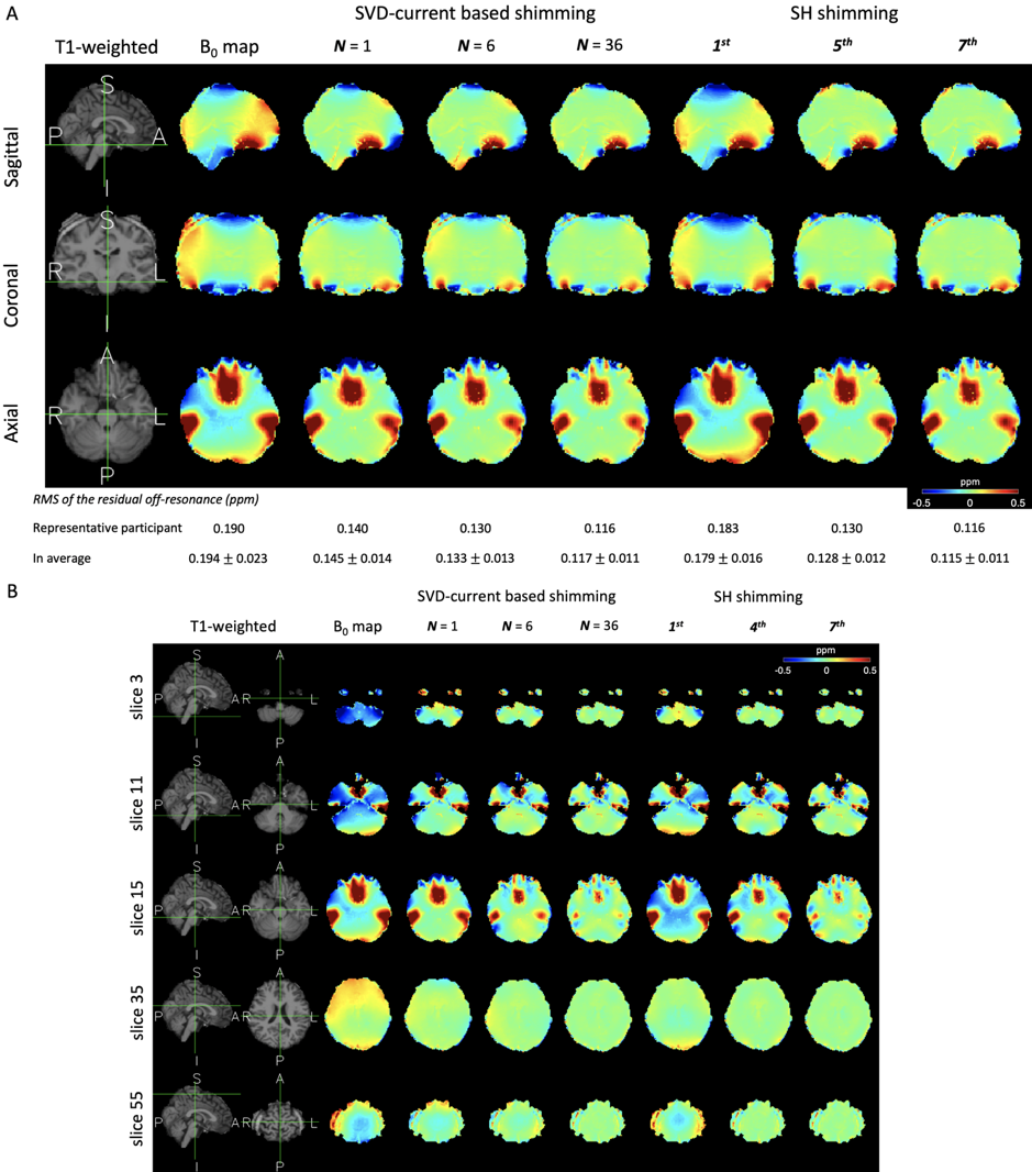
Figure 1 shows shim arrays and average as well as standard deviation of the shim current distributions across all participants. More shim coils gave more spatially smooth shim current distributions, where strong shim currents and large variations were estimated at both sides, front, and top of the head. SVD decomposed the shim currents into multiple orthogonal current modes. Figure 2 shows the average and the standard deviation of the accumulated percentage power of current modes. More than 90% of power was provided by the first 6 modes. No clear difference between shim arrays with coils distributed over a complete or a partial sphere. Figure 3 shows the first 6 current modes and the shim fields of the array with 891 circular shim coils distributed over a partial sphere. The first mode targeted bilateral temporal lobe and the orbitofrontal cortex, typical regions showing strong off resonance10,11. Higher modes targeted at other regions. Figure 4 shows the average of RMS of the residual off-resonance across 37 leave-one-out validation trials after multi-coil shimming using current modes accumulatively from different shim arrays. We noticed a transition of the shimming performance around the 6th current mode. For global shimming, using up to the 6th current mode had off-resonance about 0.13 ppm, comparable to the performance of the 5th-order SH global shimming. For slice-selective shimming, using up to the 6th current modes had the performance comparable to the 4th-order SH slice-selective shimming. Figure 5 shows the residual off-resonance distributions. The off-resonance around the frontal and bilateral temporal lobes was reduced.Discussion
SVD can suggest mutually orthogonal current modes, which can be beneficial to avoid coupling between each other in future realization. The first 6 current modes accounted for more than 90% of the variance of shim current distributions. Using all these 6 current modes together had comparable shimming to either the 5th-order or the 4th-order SH for global and slice-selective shimming, respectively. Our calculations can be taken as the upper limit of shimming performance, since we did not account for any errors in realizing the shim array.
Acknowledgements
This work was partially supported by Ministry of Science and Technology, Taiwan (103-2628-B-002-002-MY3, 105-2221-E-002-104), the National Health Research Institutes, Taiwan (NHRI-EX107-10727EI), and the Academy of Finland (No. 298131).References
- Golay M. J. E.Review of Scientific Instruments.1958;29:313-315.
- Romeo F. & Hoult D. I.Magn Reson Med.1984;1:44-65.
- Juchem C., Nixon T. W., McIntyre S.et al.J Magn Reson.2010;204:281-289.
- Juchem C., Green D. & de Graaf R. A.J Magn Reson.2013;236:95-104.
- Juchem C. & de Graaf R. A.Magn Reson Med.2017;78:2042-2047.
- Neuroimaging L. f. C. FreeSurfer software package, <https://surfer.nmr.mgh.harvard.edu/fswiki/FreeSurferWiki> (2007).
- Hoerl A. E. & Kennard R. W.Technometrics.1970;12:55-67.
- Sodickson D. K.Magnetic Resonance in Medicine: An Official Journal of the International Society for Magnetic Resonance in Medicine.2000;44:243-251.
- Caola M. J.Journal of Physics A: Mathematical and General.1978;11:
- Robin A. De Graaf C. J. in Magnetic Resonance Technology: Hardware and System Component Design (ed Andrew G Webb) Ch. 4, 172-185 (The Royal Society of Chemisty, 2016).
- Stockmann J. P. & Wald L. L.Neuroimage.2017;
Figures