1458
Initial experience with SPOKES EPI on a 7T Terra scanner1University of Cambridge, Cambridge, United Kingdom, 2Siemens UK, Frimley, United Kingdom
Synopsis
This abstract describes our initial experiences using the parallel transmit (pTx) mode on our recently-installed 7T Terra MRI scanner (Siemens, Germany). We acquired data from a phantom and a human volunteer with a turbo-FLASH B1-mapping sequence and an EPI sequence. We used the conventional single-channel system with a Nova 32-channel 1Tx head coil and the pTx system with a Nova 32-channel 8Tx head coil. For the parallel-transmit system, the sequences were run in both a static CP+ B1 shim mode, and with dynamic online SPOKES-2 pulse design. We report temporal signal-to-noise ratio (tSNR) statistics for each image series.
Introduction
We recently installed a 7T Terra MRI scanner in our MRI centre. In comparison to the previous Magnetom 7T system, the Terra comes equipped with a combined Measurement and Reconstruction Server (MARS) and runs the new VE11U software. This means that a parallel transmit (pTx) pulse1 can in theory now be implemented with negligible sequence modifications, with the scanner automatically acquiring B0 and per-channel B1-maps for adjustments, running an online Matlab-based dynamic pulse computation immediately before the measurement, and performing virtual observation points (VOP) local SAR supervision. In this abstract, we aimed to test this new framework by implementing an echo planar imaging (EPI)2 pulse sequence with the option of SPOKES-2 excitation,3-5 and comparing it against sinc excitation with a fixed CP+ B1 shim solution, and against scanning with sinc excitation on a single-channel transmit coil in the scanner’s single-channel mode.Materials and methods
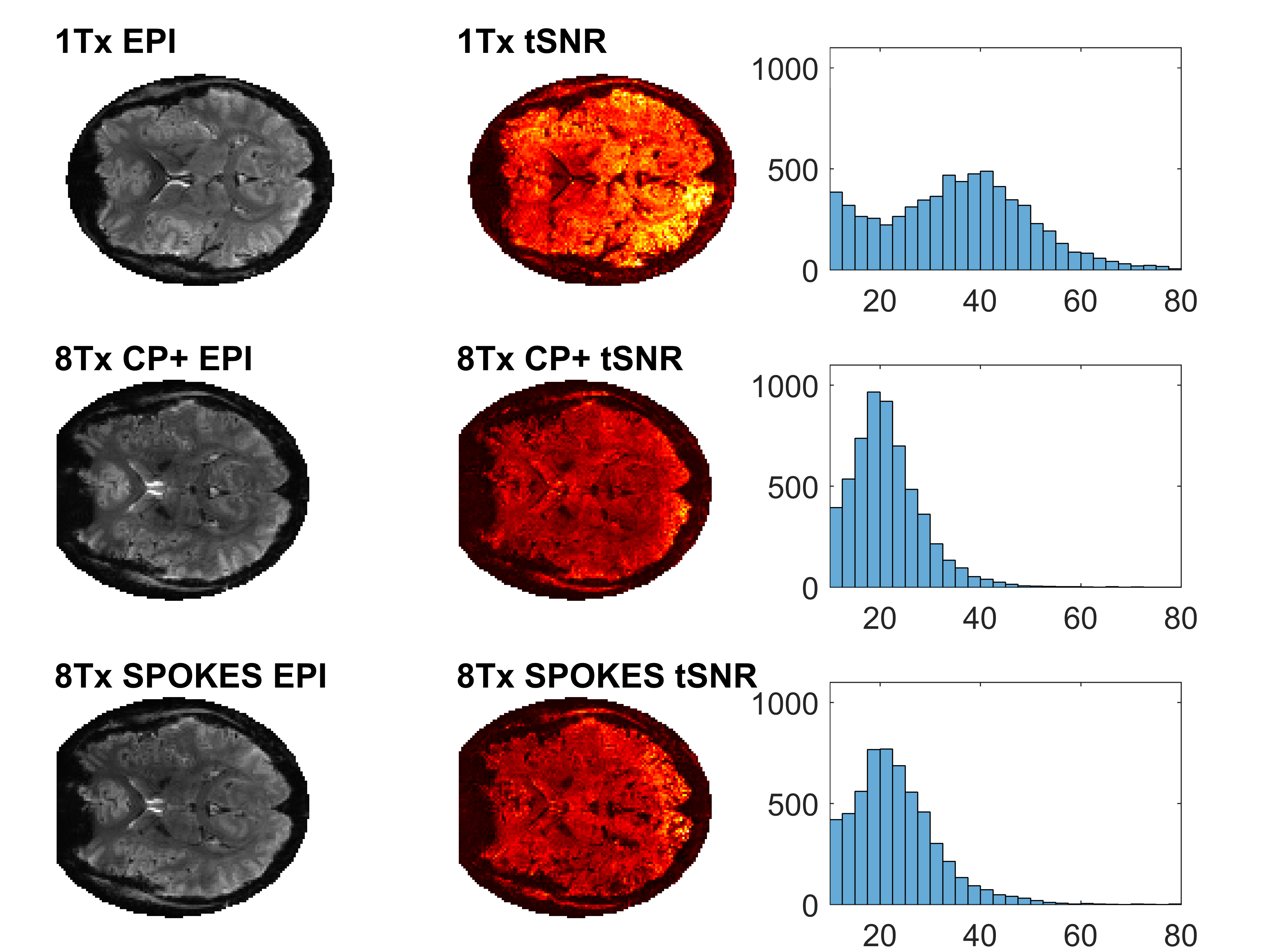
The product ep2d_bold EPI pulse sequence2 was modified to enable selection of excitation with the conventional sinc pulse, or with a dynamically computed SPOKES-2 pulse. On the pTx system, we first scanned a spherical agarose phantom.6 We acquired B1+ maps using a Turbo-FLASH B1-mapping pulse sequence (tfl_rfmap) with a static CP+ B1 shim, and a dynamically designed SPOKES-2 pulse. For both protocols, images were acquired with a matrix size of 128 × 128 over a field of view (FOV) of 220 mm × 220 mm and a slice thickness of 2.5 mm. The TR and TE were 4000 ms and 1.85 ms respectively with a flip angle of 60°. The image acquisition bandwidth was 450 Hz/Px and GRAPPA (R = 3, 24 reference lines) was used. In the CP+ mode, B1 shimming was set to ‘TrueForm’. A relative B1 map was also obtained in the static CP+ mode to obtain the single channel B1+ maps for each of the 8 channels. We then acquired 100 volumes of EPI for a single transverse slice again using CP+ B1 shim, and the dynamically designed SPOKES-2 pulse. The following parameters were used: TR = 3000 ms; TE = 25 ms; FA = 60° and bandwidth = 1562 Hz/Px to give an echo spacing of 0.74 ms. All other parameters were identical to the sequence above. Subsequently, we ran both sequences with matching parameters on the single-channel system with the ‘Advanced’ B0 shim mode. Flip angle maps were compared across all 3 protocols and tSNR statistics were analysed for each EPI series. We then scanned a healthy volunteer using a matched protocol in a single axial slice across the brain to compare the in vivo performance of the 3 modes.Results
Figure 1 shows flip angle maps in the phantom, acquired using the Turbo-FLASH B1-mapping sequence. The single-channel and CP+ mode flip angle profiles are very similar, as is expected for the two Nova coils which are designed to be as equivalent as possible. The SPOKES-2 pulse achieves a more homogeneous excitation, largely suppressing the central brightening artefact visible in the other maps. Figure 2 shows the equivalent results in vivo, and similar behaviour can be observed. Figure 3 shows the single-channel relative B1+ maps plotted for the volunteer. Figure 4 shows the EPI results for the phantom and Figure 5 shows EPI results in vivo. These show a successful implementation of dynamically designed SPOKES-2 pulse in the 2D EPI sequence. In both cases, SPOKES excitation improves the image homogeneity (left column). The changes in tSNR are less clear-cut. SPOKES increases tSNR compared to CP+ excitation, but the combined-mode single-channel coil data has the highest tSNR.Discussion and conclusions
This preliminary study has allowed us to gain hands-on practical experience implementing a pTx pulse sequence on the 7T Terra scanner. We have demonstrated that it is possible to use the new workflow for scanning phantoms and normal volunteers. The image quality for EPI improves visibly on moving from CP+ static B1-shimming to SPOKES-2 excitation. The temporal SNR shows a similar improvement. However, there appears to be an as-yet undiagnosed factor reducing the tSNR for the 8ch coil data compared to the 1ch coil data overall.
We are now ready to optimise the pTx pulse design further to allow a full stack of slices to be acquired, and to assess the fMRI performance with visual paradigms. Our 7T scanner is embedded in a tertiary referral hospital, directly adjacent to the neuro critical care ward. We are keen to discover whether pTx workflows can be made feasible for routine patient scanning in our varied population of participants.
Acknowledgements
Funded by a Sir Henry Dale Fellowship from the Wellcome Trust and the Royal Society [098436/Z/12/B] and a Gates Cambridge Scholarship to YBD. We acknowledge assistance implementing the parallel transmit workflow from our Siemens site scientist, Denis Kokorin.References
1. J. Pauly, J Magn Reson, 82, p. 571, 1989
2. P. Mansfield, J. Phys C 10:L5-L58, 1977.
3. Setsompop K, Alagappan V, Gagoski B, Witzel T, Polimeni J, Potthast A, Hebrank F, Fontius U, Schmitt F, Wald LL, Adalsteinsson E. Slice-selective RF pulses for in vivo B1+ inhomogeneity mitigation at 7 tesla using parallel RF excitation with a 16-element coil. Magn Reson Med. 2008;60(6):1422–1432.
4. Saekho S, Yip CY, Noll DC, Boada FE, Stenger VA. Fast-kz three-dimensional tailored radiofrequency pulse for reduced B1 inhomogeneity. Magn Reson Med. 2006;55(4):719–724.
5. Zelinski AC, Wald LL, Setsompop K, Alagappan V, Gagoski BA, Goyal VK, Adalsteinsson E. Fast slice-selective radio-frequency excitation pulses for mitigating B+1 inhomogeneity in the human brain at 7 Tesla. Magn Reson Med. 2008;59(6):1355–1364.
6. L. Friedman & G.H. Glover, “Report on a Multicenter fMRI Quality Assurance Protocol”, J Magn Reson Imaging. 2006 Jun;23(6):827-39.
Figures