1454
Combined volume T/R and surface Rx-only coils for simultaneous brain and spinal cord imaging of squirrel monkey at 9.4 T1Institute of Imaging Science, Vanderbilt University, Nashville, TN, United States, 2Radiology and Radiological Science, Vanderbilt University, Nashville, TN, United States
Synopsis
Simultaneous imaging of
Introduction
Functional and structural MRI play important roles in understanding the relationships between the organization of spinal cord and brain. For example, the processing of innocuous touch and painful stimuli engages circuits within the grey matters of both spinal cord and brain’s somatosensory system. These spinal cord and brain regions interact constantly to execute behavioral demands originated in the brain’s high cognitive areas such as goal-oriented limb movement. Simultaneous imaging of these separate regions potentially provides valuable information about how they work together and visualizes their interactions. However, to date, almost all image studies have investigated these two highly interconnected systems in separations, due mainly to the lack of an adequate imaging hardware. head volume coils used for brain imaging do not perform well for the spine, while surface coil arrays designed for the cord fail to image the cortex. To achieve optimal signal-to-noise-ratio for simultaneous brain and spine imaging, a combined dual coil has been designed that is suited for the smaller bore and lower number of channels typically available on high field animal MRI scanners.Methods
Figure 1a shows a conventional design typical of human systems using a volume Tx-only coil and a receive-only array1,2. However, due to the limited receive channels and space, it is hard to place such an Rx array around a monkey brain in a smaller bore 9.4T magnet. We therefore propose to use a volume coil for reception and transmission and to add a receive coil focused on the spinal cord, as shown in Figure 1b.
Figures 1c-e shows the dual coil design and how they interface to the spectrometer. The volume coil is based on the classic Alderman-Grant Resonator3 (10-cm length and 8.9-cm diameter) which can cover the whole brain and upper spinal cord of a squirrel monkey. The surface coil is a saddle-shaped loop coil with 3-cm length and 4.5-cm diameter. Figure 1f shows a schematic of the home-built T/R switch circuit. During transmission, the PIN diodes in the T/R switch (D1, D2 and D3 in Figure 2d) are ON and the transmit power goes into the volume coil. Meanwhile, the surface coil is detuned to avoid transmit field distortion (D1 in Figure 1e is ON). During reception, all PIN diodes are OFF and the MR signal will go into both coils. The two coils are carefully positioned to generate orthogonal magnetic fields and thus intrinsically isolated. To avoid cross-talk with the outer environment, a copper foiled tube (separated by 330 pF capacitors) with a diameter of 11 cm was used as shielding.
This design was validated through both EM simulation (using commercial software Ansys HFSS, Canonsburg, PA, USA) and MR experiments (9.4T Varian/Agilent small animal scanner, 12-cm bore size inside the gradients). Low flip angle GRE images with the following parameters were acquired for SNR calculations: FOV=100x100 mm2, matrix= 128x128, TR/TE=200/4 ms. Flip angle =15o, slice number=24, slice thickness/gap=2/0 mm and bandwidth=390.6 Hz/pixel.
Results and Dicussions
Figure 2a shows the simulation results. Figures 2b and 2c show the calculated vector magnetic field in an axial slice from each coil. The two coils have an orthogonal magnetic field (one horizontal and the other vertical) and are expected to be intrinsically isolated. Note the volume coil was designed to be driven linearly to work with the surface coil simultaneously during reception, which leads to ~30% SNR loss in the brain area compared to a quadrature birdcage coil.
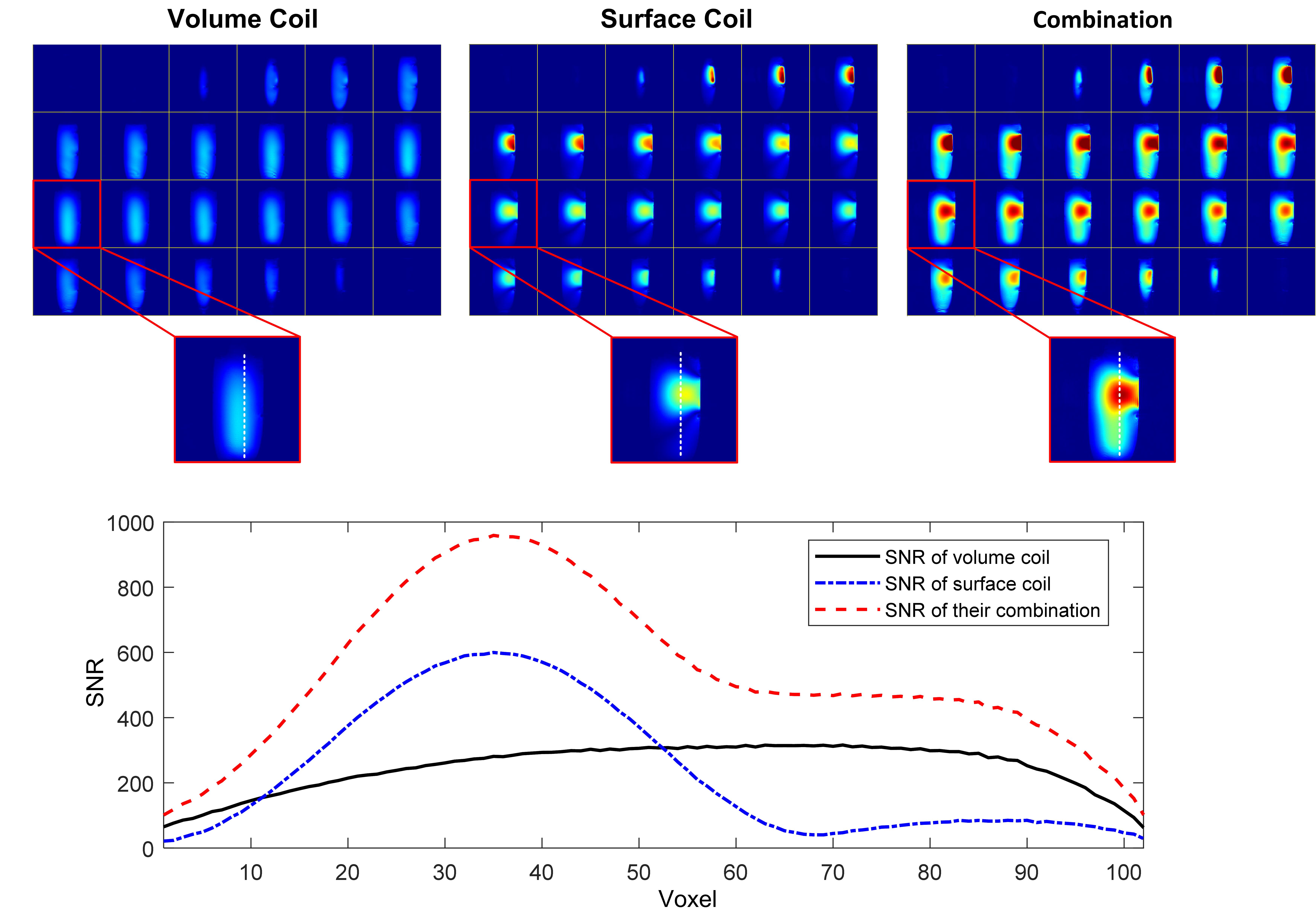
Figure 3a shows the coil built for monkey studies. Float trap circuits were used for both coils to suppress the common-mode current. The measured unloaded quality (Q-) factors of the volume coil and surface coil is 76 and 183, respectively, and their Q-factors drop to 34 and 24 when loaded with a saline phantom (4.5-cm diameter, 4g/ml NaCl). Similar to the simulation predictions, the two coils are well decoupled with S21=-27 dB (Figure 3b). Figures 4 and 5 show the measured SNR maps on this water phantom with a surface-only coil, a volume-only coil, and their combination. By using the combined coil, the SNR near the surface coil (corresponding to the spinal cord area) can be boosted by approximate 2.5 times while the SNR in other areas can be maintained.
Conclusion
We designed and fabricated a volume T/R and surface Rx-only coil for simultaneous brain and spinal cord imaging of squirrel monkey which can significantly increase the SNR around the spinal cord while simultaneously acquiring whole brain images. It needs only two receive channels and operates in a highly restricted volume, making it suitable for the small animal scanner.Acknowledgements
No acknowledgement found.References
1. Cohen‐Adad, J., et al. "32‐Channel RF coil optimized for brain and cervical spinal cord at 3 T." Magnetic resonance in medicine 66.4 (2011): 1198-1208.
2. Matschl V, Reykowski A, Jahns K, Hergt M, Fischer H. 48 Channel Body / Spine Matrix Coils for 3 Tesla; Proceedings of the 13th Annual Meeting of ISMRM; 2005.p. 952.
3. Alderman, Donald W., and David M. Grant. "An efficient decoupler coil design which reduces heating in conductive samples in superconducting spectrometers." Journal of Magnetic Resonance (1969) 36.3 (1979): 447-451.
Figures