1436
Characterization of health and technological risks posed by the quench of a human high-field Magnetic Resonance system1CIMeC, University of Trento, Trento, Italy, 2Dept of Physics, University of Trento, Trento, Italy
Synopsis
The programmed quench of a human 4T MR scanner was used to measure dB/dt inside the bore to evaluate cardiac stimulation risks during a quench. Additionally, we measured the exit temperature of the helium gas, to evaluate potential implications in quench pipe design. The maximum dB/dt was 360 mTs−1 at the center of the magnet, far below the cardiac stimulation threshold (20 Ts−1). Helium exit temperature reached 35°K, perhaps implying further considerations about quench pipe design and building. Replication of similar experiments on programmed quenches, specially in high-field MRI systems, will be useful to further characterize quench risks.
Introduction
The quench of a superconductive magnetic resonance (MR) scanner, induced or accidental, results in the fast evaporation of the liquid helium through the so-called quench pipe. It is well known that the quench pipe must be built to prevent the leakage of helium gas, and that its terminal must be positioned such that in the event of a quench there is no risk to personnel from the low temperature and high pressure helium gas. However, little is known about the temperature at which the gas can flow inside the pipe, and whether such temperature can pose any threat to the pipe itself. Another quench risk relates to the strong temporal derivative of the static field that a patient might experience if lying inside the scanner during a quench (B0 goes to the earth magnetic field in seconds). Fast magnetic field variations may induce peripheral nerve stimulation and cardiac stimulation1. Regulatory bodies have different approaches to address this risk during a quench, sometimes deeming it as negligible2, sometimes warning of possible cardiac stimulation3. However, to the best of our knowledge, there are no reports characterizing the static field derivative inside a human MR system during a quench. As part of the decommissioning of an MRI system (Bruker Medspec4T), we used the opportunity of an induced quench to measure the following parameters during the quench: i) the field changes inside the bore to assess the risk of peripheral nerve and cardiac stimulation effects, and ii) the temperature of the gas exhausted by the quench pipe.Methods
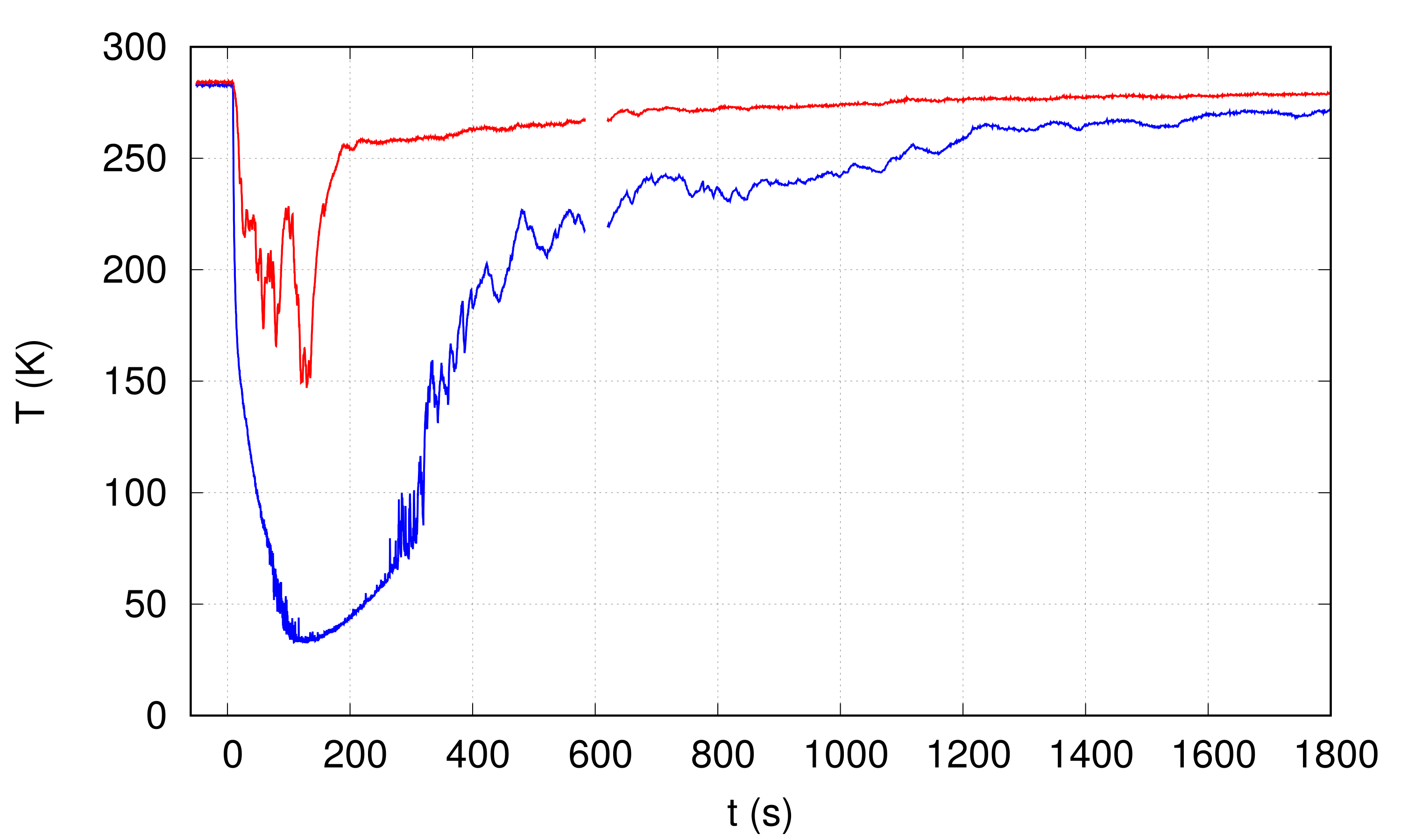
To measure the time–derivative of the field component parallel to the z–axis, a setup consisting of two pairs of coaxial coils was assembled. The axis of the coils setup coincided (within 1 cm) with the axis of the MR scanner bore. A first coil pair was placed at the bore entrance, while the other pair was placed at the center of the bore, where the magnetic field is most uniform. The temperature close to the Helium outflow pipe was measured by using two Pt100 resistors. The two sensors were placed at a distance of 10 cm and 110 cm from the output of the Helium pipe. In addition, a thermal camera was pointed to the helium exhaust pipe.Results
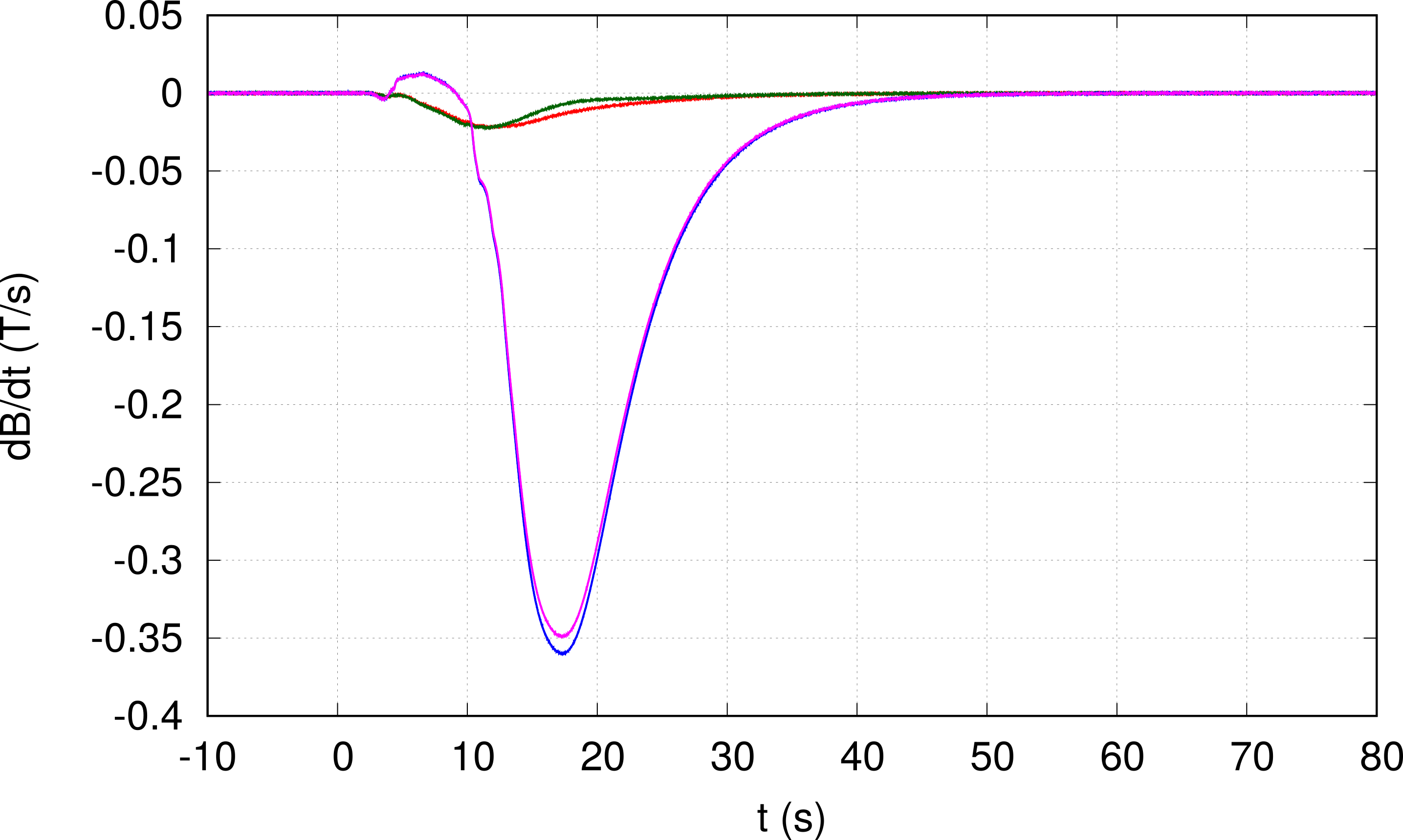
Figure 1 shows the measurements of dBz/dt for the four coils. The extinction of the magnetic field occurs in about 40 s. The maximum absolute value of dBz/dt was reached 17.0 +/- 0.5 s after the quench was initiated and was 360 mTs−1 at the center of the magnet. The coils outside the bore showed a lower maximum field derivative, 22 mTs−1.
After approximately 100 s of the quench start, the temperature 10 cm outside the quench pipe reached values as low as 35(2)°K, and remained below 50°K for almost 2 minutes. The temperature drop was less marked at 110 cm, where values as low as 150°K were reached though with a very variable trend. Environmental temperature was recovered about 3 minutes after the quench.
Discussion
According to the IEC2, the thresholds for peripheral nerve and cardiac stimulation depend upon the duration of the stimulus; after some hundreds of milliseconds, such thresholds asymptotically tends to 20 Ts-1. However, the dBz/dt values measured in this study are more than one order of magnitude lower than such threshold. Given the dynamics of an induced quench and the magnitude of the difference between our results and the cardiac stimulation threshold, it is unlikely that other human MRI scanners (with static fields equal or smaller than 4T), characterized by different manufacturing solutions, can go through a collapse of B0 fast enough to entail gradient values of the order of those needed to trigger cardiac stimulation.Our measurements of the helium temperature at the exit of the quench pipe showed that in addition to gas pressure, also gas temperature can represent a cause of potential damage for the quench pipe during the event of a quench, due to the extremely low temperature that the helium gas maintains until the exit of the pipe.Conclusions
Our data suggests that the quench of a human MR scanner like the one here tested does not seem to pose risks of cardiac stimulation in the case of a patient blocked inside the scanner. However, awareness of the extremely low temperatures reached and maintained by the helium gas all throughout the quench pipe path and at its exit is crucial to provide the best guidance and installation specification while planning a new quench pipe.Acknowledgements
No acknowledgement found.References
1. International Electrotechnical Commission (IEC). IEC 60601-2-33:2010: Medical electrical equipment - Part 2-33: Particular requirements for the basic safety and essential performance of magnetic resonance equipment for medical diagnosis, International Electrotechnical Commission (IEC), 2010
2. Medicines and Healthcare products Regulatory Agency (MHRA). Magnetic resonance imaging equipment in clinical use: safety guidelines. United Kingdom: MHRA; 2014
3. National Institute for Insurance against Accidents at Work (INAIL). Indicazioni operative dell’Inail per la gestione della sicurezza e della qualità in Risonanza Magnetica. Rome, Italy: INAIL; 2015.
Figures