1428
T1ρ and T2 Relaxation Times are Sensitive to Ischemic Injury in Femoral Head Specimens from a Piglet Model of Avascular Necrosis Independent of a Freeze/Thaw Cycle1Center for Magnetic Resonance Research, University of Minnesota, Minneapolis, MN, United States, 2Department of Radiology, University of Minnesota, Minneapolis, MN, United States, 3Department of Veterinary Population Medicine, University of Minnesota, St. Paul, MN, United States, 4Department of Veterinary Clinical Sciences, University of Minnesota, St. Paul, MN, United States, 5Texas Scottish Rite Hospital, Dallas, TX, United States, 6Department of Orthopaedic Surgery, UT Southwestern Medical Center, Dallas, TX, United States
Synopsis
We investigated whether T1, T2, and T1ρ mapping can detect early ischemic injury to bone, marrow, and cartilage in an animal model of femoral head avascular necrosis. We imaged and compared six pairs of freshly-harvested ischemic and control femoral head specimens. We then imaged the specimens a second time after a freeze/thaw cycle. We found that T1ρ and T2 mapping were sensitive to ischemic injury to the femoral heads 48 hours after onset of ischemia. Furthermore, this sensitivity was maintained after the freeze/thaw cycle. This work indicates that T1ρ and T2 mapping may help assess ischemic bone and joint disorders.
Purpose
Avascular necrosis (AVN) of the femoral head is a serious condition affecting children and young adults that can lead to severe femoral head deformity and osteoarthritis [1,2]. It is important to detect and treat AVN early to prevent deformity [1,2]. Contrast-enhanced MRI is the leading approach to detect femoral head ischemia [3], but its clinical use is limited due to safety concerns and its inability to directly detect and quantify ischemic injury to tissue. A recent ex vivo MRI study comparing quantitative mapping of T1, T2, and T1ρ relaxation times in a piglet model of AVN demonstrated that T1ρ mapping may be a particularly sensitive technique to detect ischemic injury to the femoral head as early as 48 hours after onset of ischemia [4]. A limitation of this prior study was that specimens underwent a freeze/thaw cycle before imaging. The purpose of our current study was to further investigate the sensitivity of T1, T2, and T1ρ mapping to early ischemic injury to the femoral head in the piglet model by: (i) imaging freshly-harvested femoral head specimens 48 hours after onset of ischemia; and (ii) measuring the effect of a freeze/thaw cycle on the findings. We hypothesized that: (i) T1ρ mapping would be most sensitive in detecting ischemic injury to the freshly-harvested femoral heads; and (ii) the sensitivity of the relaxation times would not be affected by a freeze/thaw cycle.Methods
Animals. Whole right femoral head ischemia was surgically induced in N=6 six-week-old male piglets by placing a tight ligature around the femoral neck and transecting the ligamentum teres [5]. The piglets were euthanized 48 hours after surgery and the operated (right) and control (left) femoral heads were harvested and refrigerated for <12 hours at 4°C prior to imaging.
Imaging. The freshly-harvested specimens were individually imaged using a preclinical 9.4T MRI scanner. The specimens were mounted to a holder and immersed in Fomblin. T1, T2, and T1ρ maps were acquired using a magnetization-prepared 2D fast spin echo (FSE) sequence (Table 1). Immediately after imaging, the specimens were wrapped in saline-soaked gauze and frozen at -20°C for 3-41 days. The specimens were then thawed at 4°C and imaged a second time using the same protocol and slice positioning.
Histology. After imaging, the thawed specimens were processed for histological analysis with H&E staining in the imaging plane. The slides were qualitatively assessed by a blinded pathologist.
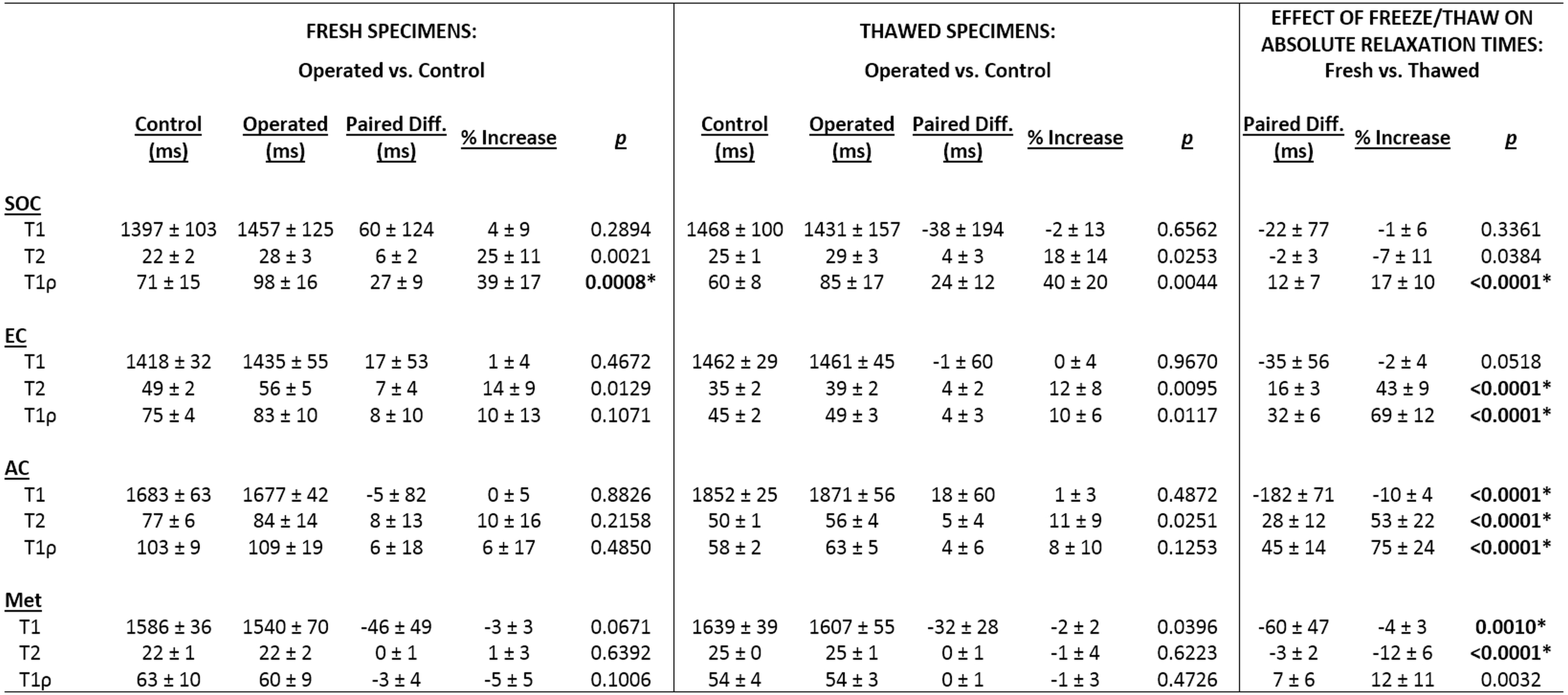
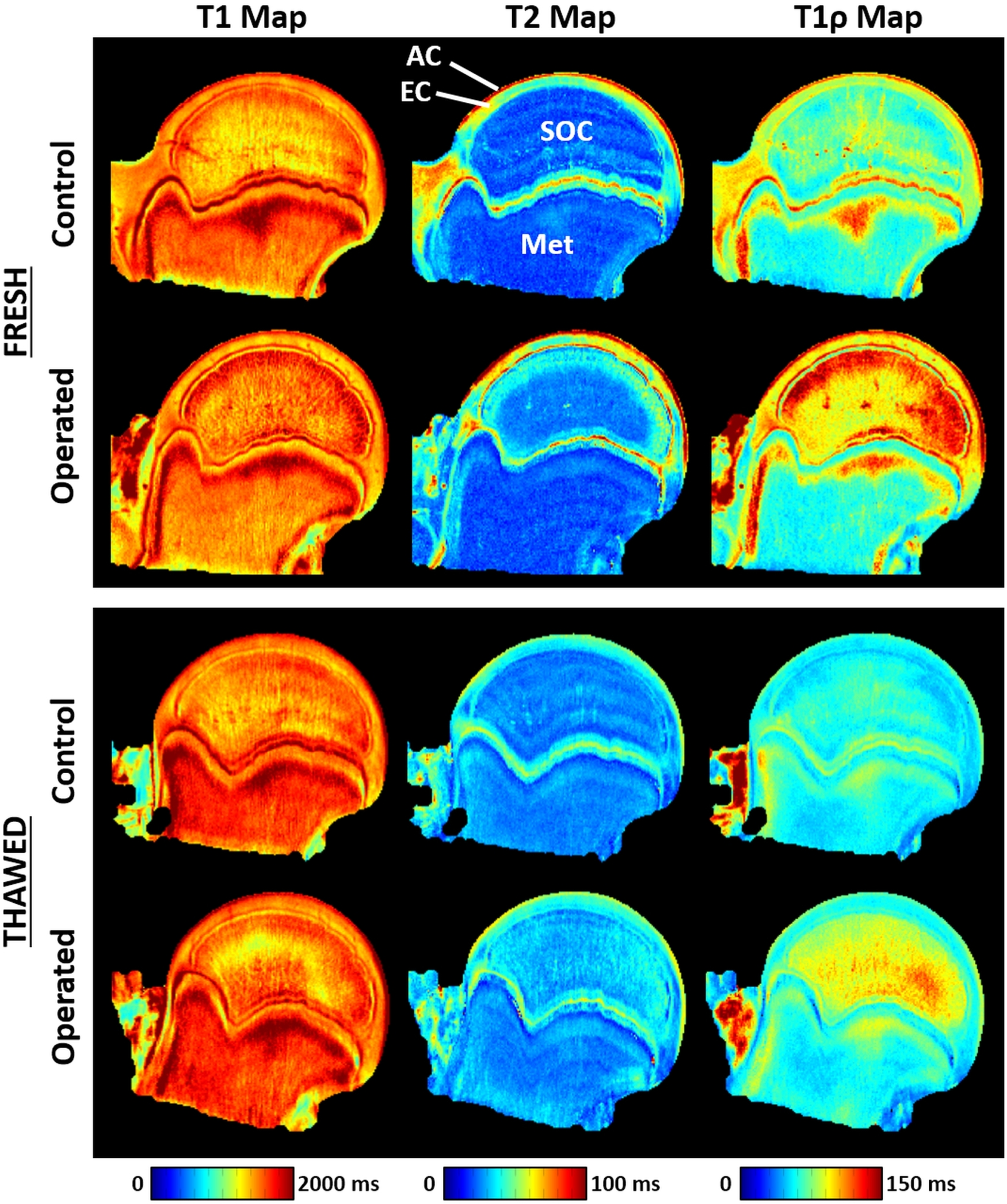
Data Analysis. Median T1, T2, and T1ρ relaxation times were calculated in four regions of interest (ROIs) (see labels in Figure 1): (i) secondary ossification center (SOC) (i.e., femoral head bone and marrow); (ii) epiphyseal cartilage (EC); (iii) articular cartilage (AC); and (iv) metaphyseal bone and marrow (Met). We tested whether the ROI measures differed between the pairs of: (i) operated and control femoral heads (both before and after freezing); and (ii) fresh and thawed specimens. Paired t-tests were used with p<0.0014 considered significant after Bonferroni correction for 36 comparisons (4 ROIs × 3 relaxation times × 3 comparisons).
Results
Histological evaluation identified extensive necrosis of the bone marrow and deep epiphyseal cartilage in 4/6 operated femoral heads, while 1/6 had subtle necrosis and 1/6 had no necrosis due to surgical failure. All femoral heads were included in the data analyses. Relaxation time maps from a piglet with extensive necrosis are shown in Figure 1, and corresponding photomicrographs are shown in Figure 2. Results of the ROI analyses for all six piglets are tabulated in Table 2. The freeze/thaw cycle resulted in a significant decrease in T1ρ relaxation times in the SOC, EC, and AC. T2 relaxation times were also significantly decreased in the EC and AC. Despite these changes in absolute relaxation times, the percent increase in relaxation times between the operated and control femoral heads were remarkably similar before and after the freeze/thaw cycle (Figure 3). T1ρ had the greatest percent increase in the SOC due to ischemic injury (~40%), which was maintained after freezing.Discussion
Our findings support that: (i) T1ρ and T2 mapping can detect early ischemic injury to the femoral head; and (ii) their sensitivities are maintained after a freeze/thaw cycle. This work motivates investigation of T1ρ and T2 mapping as non-contrast-enhanced methods for early detection of AVN in the femoral head and other regions. Furthermore, the techniques can be effectively used to detect ischemic injury after a freeze/thaw cycle, which corroborates the initial report [4] and will facilitate future investigation of T1ρ and T2 mapping sensitivities to AVN, their contrast mechanisms, and the pathophysiology of and potential treatments for AVN.Acknowledgements
This study was supported by the NIH (K01AR070894, K01OD021293, and P41EB015894), the W. M. Keck Foundation, and Texas Scottish Rite Hospital for Children.References
1. Moya-Angeler J, Gianakos AL, Villa JC, Ni A, Lane JM. Current concepts on osteonecrosis of the femoral head. World J Orthop. 2015; 6(8):590-601.
2. Larson E, Jones LC, Goodman SB, Koo KH, Cui Q. Early-stage osteonecrosis of the femoral head: where are we and where are we going in year 2018? Int Orthop. 2018; 42(7):1723-1728.
3. Jaramillo D, Villegas-Medina OL, Doty DK, Dwek JR, Ransil BJ, Mulkern RV, Shapiro F. Gadolinium-enhanced MR imaging demonstrates abduction-caused hip ischemia and its reversal in piglets. AJR Am J Roentgenol 1996; 166(4):879-87.
4. Johnson CP, Wang L, Tóth F, Aruwajoye O, Carlson CS, Kim HK, Ellermann JM. Quantitative MRI helps to detect hip ischemia: preclinical model of Legg-Calvé-Perthes disease. Radiology 2018; 289(2):386-395.
5. Kim HK, Su PH. Development of flattening and apparent fragmentation following ischemic necrosis of the capital femoral epiphysis in a piglet model. J Bone Joint Surg Am 2002; 84-A(8):1329-34.
Figures