1423
DANTE-SPACE-based imaging of the brachial plexus1MR Collaborations, Siemens Healthcare Ltd., Shenzhen, China, 2BGI, Shenzhen, China, 3Siemens Healthcare Ltd., Guangzhou, China, 4MR Collaborations, Siemens Healthcare Ltd., Shanghai, China, 5Siemens Healthcare GmbH, Erlangen, Germany, 6Department of Biomedical Engineering, School of Basic Sciences, Guangzhou Medical University, Guangzhou, China, 7Shenzhen Institutes of Advanced Technology, Shenzhen, China
Synopsis
Magnetic resonance neurography (MRN) has been increasingly used to evaluate brachial plexopathy. However, MRN is limited by the lack of relative contrast between nerves and their surrounding tissues. As an alternative to MRN, T2-SPACE has been proposed as a black-blood technique that permits more direct visualization of the brachial plexus; however, it has similar limitations. A preliminary study demonstrated that T2-weighted DANTE-SPACE outperformed SPACE and might be a potential alternative for the visualization of the brachial plexus. In this study, we hypothesized that T2-weighted DANTE-SPACE would address the above issue due to its superior blood flow suppression.
Introduction
The
brachial plexus is a network of converging and branching nerves that supply the
terminal motor and sensory branches to the upper extremities. Distinguishing
brachial plexopathy from other spine-related abnormalities often poses a
considerable diagnostic challenge due to the location of the plexus deep within
the axilla. Magnetic resonance neurography (MRN) has been increasingly used in
recent years to further evaluate cases of suspected or established brachial
plexopathy, but it is limited by the lack of relative contrast between nerves
and their surrounding tissues [1]. Recently, 3D T2-weighted variable-flip-angle
turbo spin-echo (SPACE) has been proposed as a black-blood technique that
permits more direct visualization of the brachial plexus [2]. Although SPACE
has an inherent dark blood effect due to dephasing over the turbo spin echo
train, signal suppression may not be complete for slow venous flow or after
intravenous contrast administration [2]. Furthermore, this technique has limited
capacity due to the high signal within veins and the insufficient contrast
between nerves and their surrounding tissues. We hypothesized that T2-weighted
SPACE with DANTE preparation might address the above issues due to its superior
blood flow and CSF suppression [3-5].
Methods
Experiment: This IRB-approved study was performed in 33 healthy volunteers on a 3T MAGNETOM Prisma scanner with a 20-channel head coil and an 18-channel body matrix coil (Siemens Healthcare, Erlangen, Germany). The optimized parameters for prototypic T2-weighted DANTE-SPACE with DANTE preparation included: FA=10°, pulse trains=148, RF gap=1.13ms, spoiler moment=18000(mT/m*usec). The parameters for the SPACE readout included: 3D coronal imaging with a resolution of 1.0 × 1.0 × 1.2mm3, FOV=320mm2, TR/TE=3600/206ms, turbo factor=100, GRAPPA factor=2, bandwidth=539Hz/pixel. Conventional T2-weighted SPACE with the same parameters was also conducted for comparison.Image Analysis: Using the method similar to [2], contrast-to-noise ratios (CNRs) between the nerves and their surrounding tissues were calculated for all the subjects and compared with those obtained by the T2-SPACE approach. Two experienced radiologists scored the quality of the images generated by both DANTE-SPACE and T2-SPACE on a four-point scale (4 points indicated intact, continuous, and clear visualization of the brachial plexus and its main branches without venous interference; 3 points indicated that the majority of the brachial plexus and its branches were displayed continuously and with some slight venous interference which had no impact on evaluation; 2 points indicated that the majority of the brachial plexus and its branches were displayed and clearly with some moderate venous interference which had impact on evaluation; 1 point indicated the majority of the brachial plexus structure was not displayed and there was serious venous interference). CNRs and image quality scores between the two methods were compared using t test. Statistical significance was defined as p < 0.05.
Results
In contrast to T2-SPACE, the images obtained by the proposed DANTE-SPACE method had better CNR (197.80±71.52 vs. 145.50±74.71, p = 0.005) between the nerves and their surrounding tissues. Furthermore, the proposed DANTE-SPACE method provided a superior image quality score compared to the T2-SPACE technique (3.25 ± 0.46 vs. 1.88± 0.35, p = 0.008).
Discussion
To our
knowledge, this is the first time a study has investigated the feasibility of
the DANTE-SPACE technique for brachial plexus imaging. T2-weighted DANTE-SPACE
can provide excellent venous blood signal suppression and definitive
visualization of the brachial plexus. This preliminary study has demonstrated
that the technique may outperform SPACE and potentially become an alternative
for the diagnosis of brachial plexus pathology. A large clinical study is
underway to validate the clinical value of this technique.
Conclusion
Compared to T2-SPACE, DANTE-SPACE can effectively improve the delineation of the brachial plexus by inhibiting the high signal from veins and has the potential to be used clinically for diagnosing pathology of the brachial plexus.Acknowledgements
No acknowledgement found.References
[1] Chhabra A, Thawait G K, Soldatos T, et al. High-Resolution 3T MR Neurography of the Brachial Plexus and Its Branches, with Emphasis on 3D Imaging[J]. American Journal of Neuroradiology, 2013, 34(3): 486-497.
[2] Wang L, Niu Y, Kong X, et al. The application of paramagnetic contrast-based T2 effect to 3D heavily T2W high-resolution MR imaging of the brachial plexus and its branches[J]. European Journal of Radiology, 2016, 85(3): 578-584.
[3] Xie G, Chen H, He X, et al. Black-blood thrombus imaging (BTI): a contrast-free cardiovascular magnetic resonance approach for the diagnosis of non-acute deep vein thrombosis[J]. Journal of Cardiovascular Magnetic Resonance, 2017, 19(1): 4.
[4] Chen H, He X, Xie G, et al. Cardiovascular magnetic resonance black-blood thrombus imaging for the diagnosis of acute deep vein thrombosis at 1.5 Tesla[J]. Journal of Cardiovascular Magnetic Resonance, 2018, 20(1): 42.
[5] Li L, Kong Y, Zaitsu Y, Matthews L, Palace J, and Jezzard P. Structural Imaging of the Cervical Spinal Cord with Suppressed CSF Signal Using DANTE Pulse Trains 2015. Magnetic Resonance in Medicine, Vol. 74, pp. 971–977.
Figures
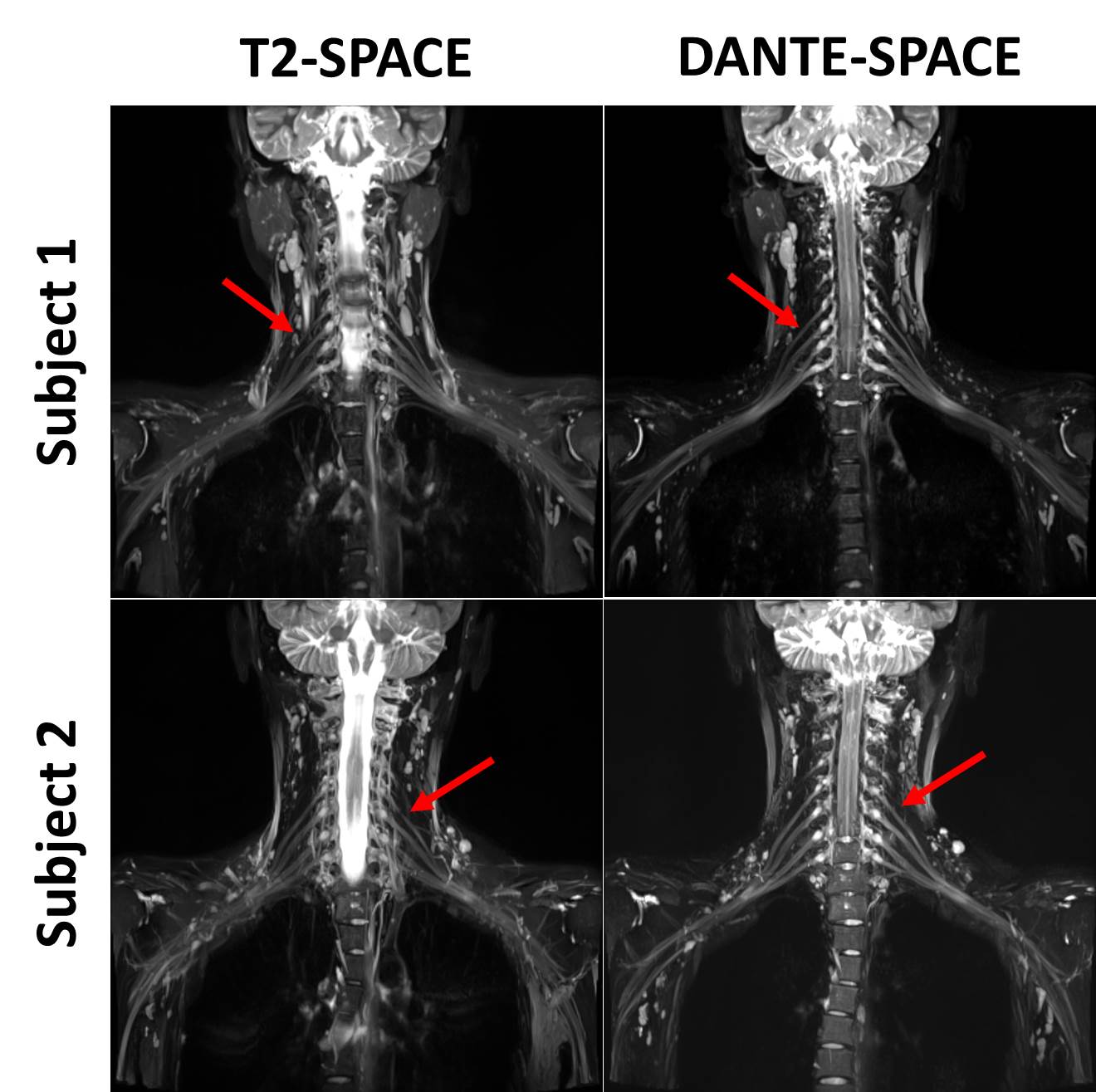
Figure.1 Representative coronal images obtained from two subjects using T2-weighted DANTE-SPACE (right column) and T2-SPACE (left column).