1422
Effects of trajectory and k-space undersampling in Compressed Sensing-Parallel Imaging 3D-GRASE1Radiology and Nuclear Medicine, Erasmus MC, Rotterdam, Netherlands, 2Medical Informatics, Erasmus MC, Rotterdam, Netherlands, 3Healthcare Systems, GE Healthcare, Hoevelaken, Netherlands
Synopsis
Compressed sensing parallel imaging (CSPI) 3D-GRASE can reduce the acquisition time compared to CSPI 3D-FSE. Image quality of 3D-GRASE strongly depends on the sampling pattern used, since gradient-echoes (GREs) and spin-echo (SE) are combined in the same k-space. Moreover, successful CSPI relies on appropiate incoherent sampling patterns.
In this work we propose and investigate the influence of several sampling patterns on coherence and in-vivo image quality of $$$PD$$$-weighted knee CSPI 3D-GRASE. With the best sampling pattern CSPI 3D-GRASE obtain high image quality with significantly reduced acquisition time (57%) and SAR (66%) compared to CSPI 3D-FSE acquisitions.
Introduction
High-resolution three-dimensional (3D) structural MRI is useful for delineating complex or small structures of the body1. However, it requires long acquisition times, limiting its clinical use. Compressed Sensing-Parallel Imaging (CSPI) 3D-FSE has recently been proposed to accelerate knee imaging2.
CSPI 3D-GRASE can reduce the acquisition time compared to CSPI 3D-FSE3. Image quality in 3D-GRASE strongly depends on the sampling pattern used, since gradient-echoes (GREs) and spin-echo (SE) are combined in the same k-space4. Moreover, successful CSPI relies on the design of an incoherent sampling pattern.
In this work we propose and investigate several sampling patterns in terms of coherence and in-vivo image quality of $$$PD$$$-weighted knee CSPI 3D-GRASE.
Methods
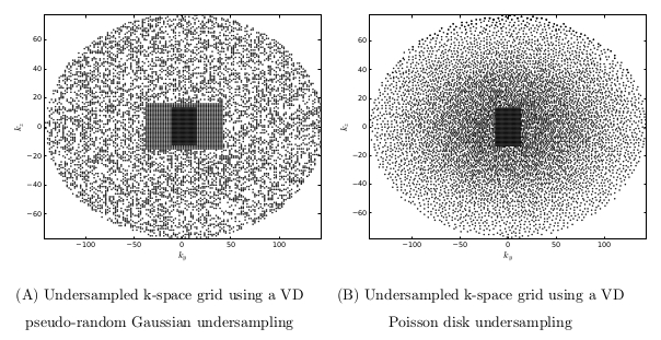
The proposed sampling patterns were based on two undersampled k-space grids, variable density Poisson-disc (VD-P) and variable density pseudo-Gaussian random (VD-G)(see Figure 1), and five different trajectories previously described in the literature (SRE1, SRE2, SLCE1, SCLE2)4. Each trajectory modulates $$$T_2$$$ and $$$T_2$$$-effects differently, which can result in artefacts.
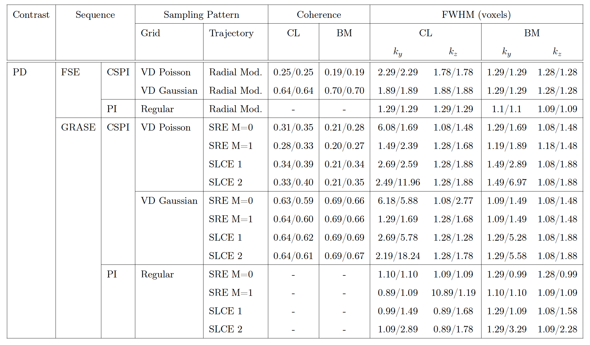
Bloch simulations were performed to obtain the transform point spread function (TPSF). Two tissues were simulated with the following properties: $$$T_1$$$=1240ms $$$T_2$$$=36.9ms $$$B_0$$$=50Hz, corresponding to cartilage (CL); and $$$T_1$$$=371ms $$$T_2$$$=133ms $$$B_0$$$=50Hz , corresponding to bone marrow (BM). The sidelobe-to-peak ratio (SPR) was obtained from the TPSF as the coherence measurement5. Moreover, the FWHM was obtained as resolution measure on the PSF for every sampling pattern.
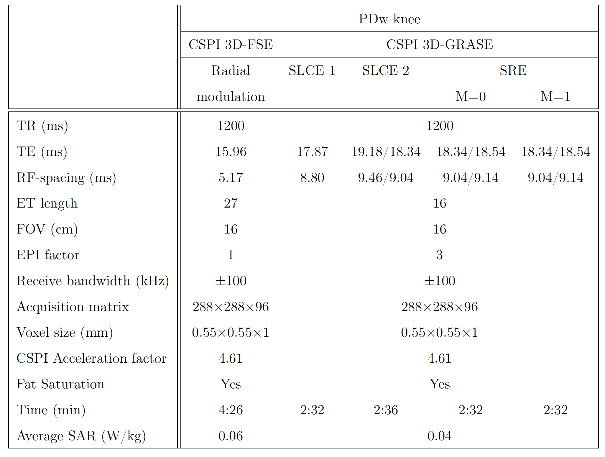
Images from two volunteers were acquired on a 3T GE Discovery MR750 (General Electric Medical Systems, Waukesha, WI) with an eight-channel phase-array transmit-receive knee coil (Precision Eight TX/TR High-Resolution Knee Array, In Vivo, FL) for knee $$$PD$$$-weighted. Protocol parameters for PI 3D-FSE, CSPI 3D-FSE, PI 3D-GRASE and CSPI 3D-GRASE are shown in Table 1. CSPI image reconstruction was performed by ESPIRit6 with l2-wavelet regularization.
To evaluate image quality in in-vivo imaging, the SNR was measured in cartilage (CL) and bone marrow (BM), as well as, the perception based image quality evaluator (PIQE)7. PIQE obtains a quality score measurement (QScore), where the lower the value, the higheer the image quality. SPR, FWHM, SNR and PIQE were obtained in Parallel-imaging (PI) 3D-GRASE and PI 3D-FSE sequences and used as gold standard.
Results
Table 2 shows the coherence and FWHM values of every sampling pattern. The coherence measure is lower with the VD-P undersampled k-space grid for all trajectories. There are more differences among the trajectories in cartilage than in bone marrow. The FWHM measurement is, in general, also smaller for VD-P trajectories. Among trajectories, the SRE1 and SRE2 show smaller FWHM along $$$k_y$$$. The FWHM is higher in CSPI than in PI simulations, which suggest higher blurring in the images.
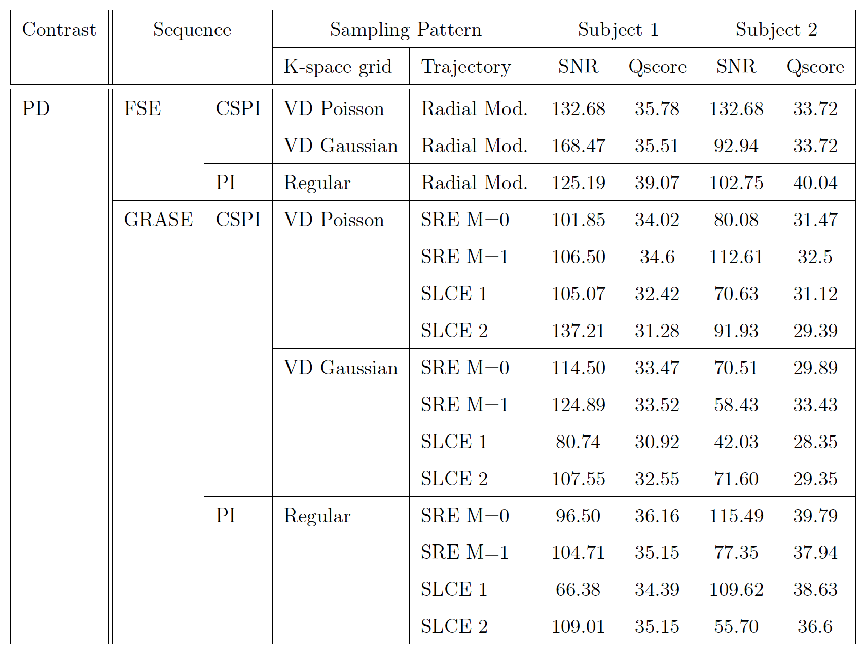
Table 3 shows the SNR and QScore values of every sampling pattern evaluated in the in-vivo acquisitions. For both volunteers, both the highest SNR and the lowest Qscore were obtained with the sampling pattern combining the SLCE 2 trajectory with the VD-P k-space grid.
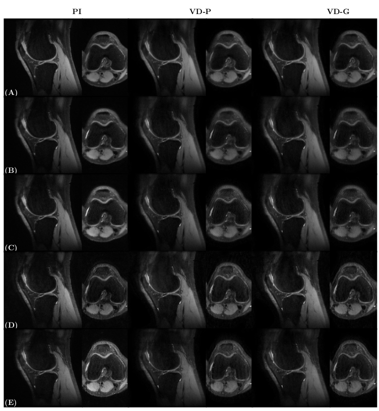
Figure 2 shows the prospective in-vivo acquisitions with each PI and CSPI sampling pattern for 3D-GRASE and 3D-FSE in one volunteer. Only the sagittal and axial planes are shown. Blurring can be appraised in some of the trajectories, especially in the axial plane. Image quality is mostly preserved for the sampling pattern combining VD-P and SLCE2, as showed by the SNR and QScore.
Discussion/Conclusions
In CSPI 3D-GRASE, image quality is preserved if the correct sampling pattern is chosen. We observed that the k-space grid is the most influencing factor for the coherence measurement, since the difference in the coherence, SNR and Qscore values among trajectories are low. Higher SNR values allow distinguishing and visualizing small structures of the knee. On the other hand, sampling patterns with lower coherence values ensure a more stable CS reconstruction. Finally, a low Qscore implies that the sampling pattern obtains images which have less distortions, based on the statements of this method.
CSPI 3D-GRASE significantly reduces acquisition time (57%) and SAR (66%) compared to CSPI 3D-FSE acquisitions. The design of the sampling pattern, including both the undersampled k-space grid and the k-space trajectory, is crucial to obtain high image quality in in-vivo CSPI 3D-GRASE.
Acknowledgements
No acknowledgement found.References
1. Yamabe E, Anavim A, Sakai T, Miyagi R, Nakamura T, Hitt D, Yoshioka H. Comparison between high-resolution isotropic three-dimensional and high-resolution conventional two-dimensional FSE MR images of the wrist at 3 tesla: A pilot study. J Magn Reson Imaging. 2014;40:603–608.
2. Pandit P, Rivoire J, King K, Li X. Accelerated T1rho acquisition for knee cartilage quantification using compressed sensing and data-driven parallel imaging: A feasibility study. Magn Reson Med. 2016;75:1256–1261.
3. Cristobal-Huerta A, Poot DHJ, Vogel M, Hernandez-Tamames JA. Compressed Sensing Variable Flip Angle 3D-GRASE for T2-weighted High-Resolution Brain Images. In Proceedings of the 34th annual scientific meeting of ESMRMB, Barcelona, 2017. p. 184.
4. Cristobal-Huerta A, Poot DH, Vogel MW, Krestin GP, Hernandez-Tamames JA. Kspace trajectories in 3D-GRASE sequence for high resolution structural imaging. Magn Reson Imaging. 2018;48:10–19.
5. Lustig M, Donoho D, Pauly JM. Sparse MRI: The application of compressed sensing for rapid MR imaging. Magn Reson Med.. 2007;58:1182–1195.
6. Lai P, Lustig M, Brau A, Vasanawala S, Beatty P, Alley M. Efficient L1-SPIRiT reconstruction (ESPIRiT) for highly accelerated 3d volumetric MRI with parallel imaging and compressed sensing. In Proceedings of the 18th Annual Meeting of ISMRM, Stockholm, Sweden, 2010. p. 345.
7. Venkatanath N, D. Praneeth, Maruthi Chandrasekhar Bh, Sumohana S.Channappayya, and Swarup S. Medasani. "Blind image quality evaluation using perception based features." In Communications(NCC), 2015 Twenty First National Conference on, pp.1-6. IEEE, 2015.
Figures