1415
MRI Off-Resonance Analysis Correlates with Histology Necrosis Observations in Failed Total Hip Arthroplasty1Radiology, Medical College of Wisconsin, Milwaukee, WI, United States, 2Biomedical Engineering, Marquette University and Medical College of Wisconsin, Milwaukee, WI, United States, 3Magnetic Resonance Imaging, Hospital for Special Surgery, New York, NY, United States
Synopsis
This study presents the analysis of 78 total hip arthroplasty revision subjects that underwent advanced MRI off-resonance analysis and optical histology necrosis grading of tissue biopsies. The off-resonance analysis, which leverages multi-spectral MRI methods, sought to quantify metal particle deposition in tissues or “metallosis”. Direct measurements of off-resonance signatures correlated strongly with histology necrosis grade (p < 0.013). The results of this study provide encouragement that quantitative MRI may be useful as a non-invasive biomarker of tissue destruction in symptomatic total hip arthroplasty.
Introduction
One in every one-hundred American adults has an artificial hip joint. All hip arthroplasties have inevitable wear-induced and/or inflammatory failure modes [1, 2, 3], which depend heavily on patient-specific risk and genetic factors. Post-surgical pain is eventually reported in as many as 40% of patients after hip arthroplasty [4]. Of the over 400,000 hip arthroplasty (HA) surgical procedures performed annually in the United States, nearly 15% are revision procedures that result in partial or complete removal and replacement of the prosthesis [5, 6].
Indirect observation of substantial metallic debris on MRI in peri-implant tissues can confirm the generation of wear debris at different articulations within an arthroplasty design. Metallic debris deposition into the surrounding soft tissue envelope, which is often described as “metallosis,” leads to tissue necrosis of surrounding structures, such as hip abductors, resulting in patient morbidity with altered gait mechanics and pain.
Recently, a method has been presented that allows MRI to be utilized to quantify off-resonance signatures from metal particle deposits near total hip replacements [7]. An initial demonstration of this technology analyzed a small (N=27) cohort of failed total hip replacements and found a significant correlation with regions of suspected metallosis. This initial study also identified a preliminary trend correlating MRI-based off-resonance and histological necrosis grading. In the current study, we performed MRI off-resonance analysis on a larger cohort of failed hip replacements (N=78) and performed a detailed comparison with recorded histological necrosis grades.
Methods
This study was approved by the local Institutional Review Board, and all subjects provided written informed consent prior to participating in the study. MRI off-resonance analysis was performed on 78 subjects undergoing revision for THA.
Prior to revision surgery, enrolled subjects underwent an MRI exam including MAVRIC SeLective (SL) 3D-MSI. Image acquisition parameters were as follows: coronal scan plane, 36–40 cm field of view, 7 ms echo time, 4 s repetition time, echo train length of 20, 5 mm slice thickness, (512x256x24–32 data matrix, 24 spectral bins, spectral width of 2.25 kHz.
For each subject, a region of suspected metallosis was identified on a MAVRIC SL image by radiologists on the study team. This regional identification was used to guide extraction of a 1 cm^3 tissue sample during the surgical revision procedure. These regions were denoted as areas of low signal intensity within the synovial envelope on the MAVRIC SL images. Extracted samples were fixed in formalin and processed for routine histology; representative sections were then graded by a board-certified pathologist.
Off resonance analysis was performed following the algorithms presented in [7]. Briefly, the following procedure was implemented.
1) Construction of 3D-MSI off-resonance map [8]
2) Regional selection of 120cm3 volume around indicated tissue biopsy region
3) Background removal using conventional QSM approaches [9]
4) Computation of “mScore” using cluster-based off-resonance analysis [7]
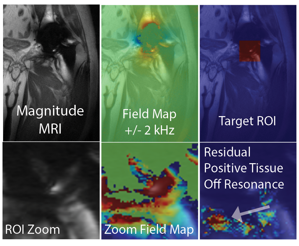
An illustration of imaging results from each step of this computational procedure is provided in Figure 1. Linear regression analysis was performed using the necrosis histology grades (1 through 4), computed mScores, and positive off-resonance voxel-wise averaging within the ROI.
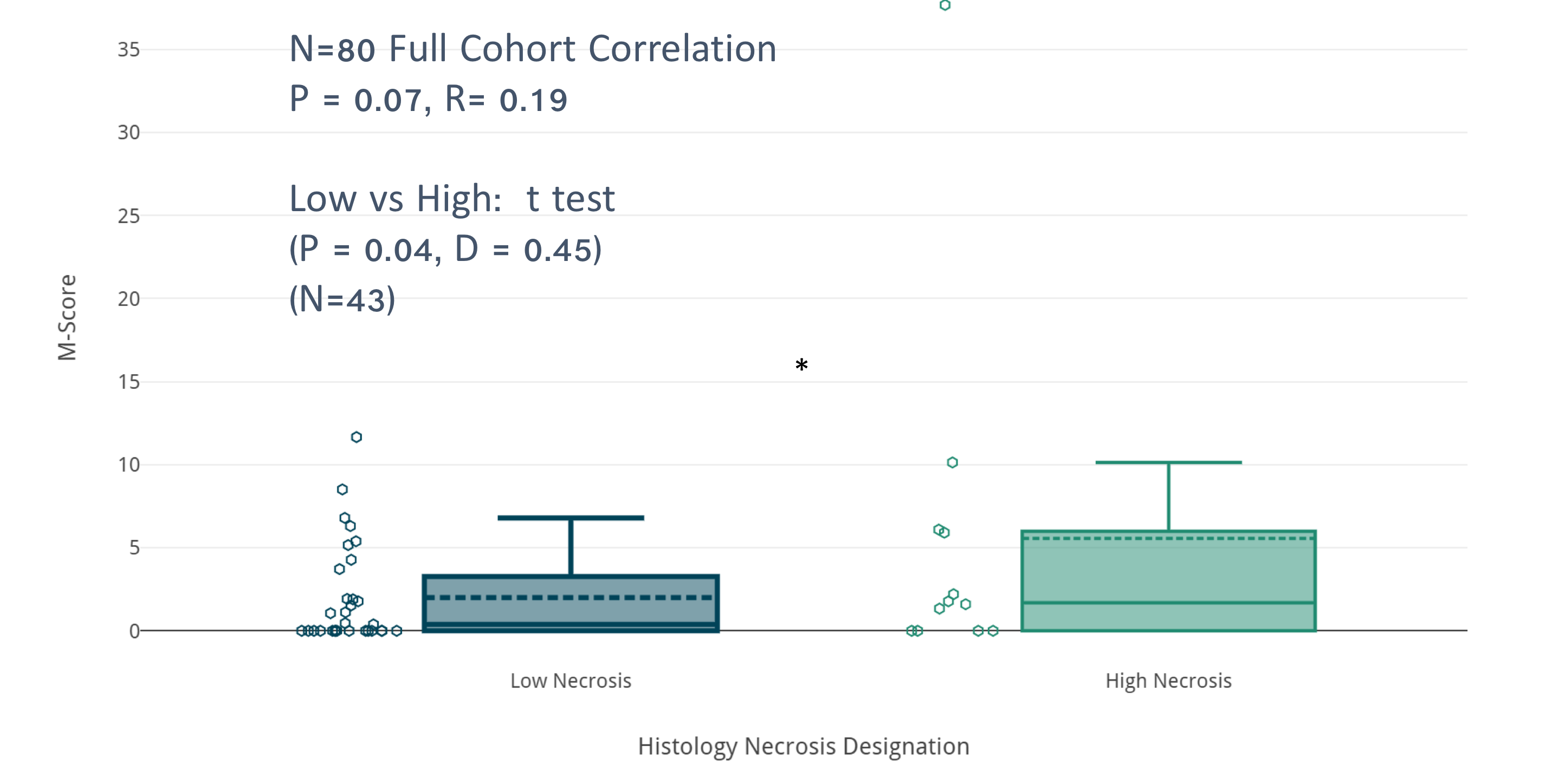
In addition, a student t-test was performed on mScores within the groups classified by low (1) and (4) histology necrosis grades.
Results
ANOVA regression showed that computed mScores correlated with necrosis grade at a slightly higher level to that identified in the smaller cohort analysis (p = 0.07, R=0.2). However, an extremum analysis at the endpoints of the histology grade (1 & 4) showed a stronger distinction of mScore classification (p = 0.04) (Figure 2).
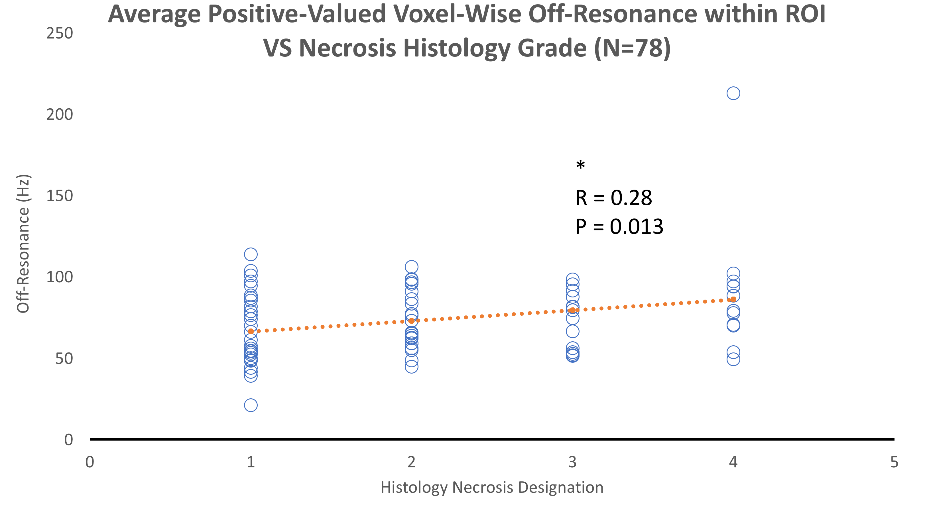
After computing off-resonance signatures based on an average of all positive off-resonance voxels within each ROI, regression analysis showed a significant correlation with necrosis grade (P=0.013, R=0.28). Scatter plots of this correlation are shown in Figure 3.
Discussion
The previously described mScore approach [7] utilized cluster analysis and off-resonance thresholds to avoid false positive classification in regions within suspected metallosis. This approach was applied to the current study and did not show any false positives in control regions extracted from each of the 78 subjects analyzed in the present cohort.
However, the results of this study show that the mScore approach reduces sensitivity to necrosis signatures. Using a direct assessment of off-resonance estimates, the correlation with optical histology necrosis grading was substantially improved and demonstrated a clear statistically significant correlation.
Further work will investigate improved methods off-resonance quantification, including quantitative susceptibility mapping approaches. Leveraging a non-invasive MRI biomarker to predict necrosis progression could have substantial impact on management of failed hip arthroplasty. The present study provides further evidence that such biomarkers can be derived from MRI data.
Acknowledgements
This work was funded in part by a technology development grant from GE Healthcare.
Research reported in this publication was supported by NIH/NIAMS R01AR064840. The content is solely the responsibility of the authors and does not necessarily represent the official views of the NIH.
References
[1] H. Maradit Kremers, D. R. Larson, C. S. Crowson, W. K. Kremers, R. E. Washington, C. A. Steiner, W. A. Jiranek, and D. J. Berry. Prevalence of Total Hip and Knee Replacement in the United States. J. Bone Joint Surg. Am., 97(17):1386–1397, September 2015.
[2] H. C. Amstutz, P. Campbell, N. Kossovsky, and I. C. Clarke. Mechanism and clinical significance of wear debris-induced osteolysis. Clinical orthopaedics and related research, 276:7–18, 1992.
[3] J. Hauptfleisch, H. Pandit, G. Grammatopoulos, H. S. Gill, D. W. Murray, and S. Ostlere. A MRI classification of periprosthetic soft tissue masses (pseudotumours) associated with metal-on-metal resurfacing hip arthroplasty. Skeletal radiology, 41(2):149–155, 2011.
[4] T. E. Brown, B. Larson, F. Shen, and J. T. Moskal. Thigh Pain After Cementless Total Hip Arthroplasty: Evaluation and Management. Journal of the American Academy of Orthopaedic Surgeons, 10(6):385–392, 2002.
[5] S. M. Kurtz, K. L. Ong, E. Lau, and K. J. Bozic. Impact of the Economic Downturn on Total Joint Replacement Demand in the United States. J. Bone Joint Surg. Am., 96(8):624–630, April 2014.
[6] K. J. Bozic, A. F. Kamath, K. Ong, E. Lau, S. Kurtz, V. Chan, T. P. Vail, H. Rubash, and D. J. Berry. Comparative Epidemiology of Revision Arthroplasty: Failed THA Poses Greater Clinical and Economic Burdens Than Failed TKA. Clinical Orthopaedics and Related ResearchR , 473(6):1–8, April 2015.
[7] Koch, K.M., Koff, M.F., Bauer, T.W., Shah, P.H., Nencka, A.S., Sivaram Kaushik, S. and Potter, H.G., 2018. Off‐resonance based assessment of metallic wear debris near total hip arthroplasty. Magnetic Resonance in Medicine, 79(3), pp.1628-1637.
[8] Koch, K.M., Brau, A.C., Chen, W., Gold, G.E., Hargreaves, B.A., Koff, M., McKinnon, G.C., Potter, H.G. and King, K.F., 2011. Imaging near metal with a MAVRIC‐SEMAC hybrid. Magnetic Resonance in Medicine, 65(1), pp.71-82.
[9] Liu, T., Khalidov, I., de Rochefort, L., Spincemaille, P., Liu, J., Tsiouris, A.J. and Wang, Y., 2011. A novel background field removal method for MRI using projection onto dipole fields. NMR in Biomedicine, 24(9), pp.1129-1136.
Figures