1402
Could the 3D CAIPIRINHA accelerated SPACE imaging replace the conventional 2D MRI in routine knee examination?1Radiology, Tongji Hospital, Tongji Medical College, Huazhong University of Science and Technology, Wu Han, China, 2Siemens Healthcare Ltd., Shanghai, China, Shanghai, China, 3Siemens Healthcare Ltd.,Shenzhen, China, Shen Zhen, China
Synopsis
3D CAIPIRINHA accelerated SPACE is a newly
developed technic that can achieve faster acquisition and high resolution
isotropic 3D imaging of the knee. However, its clinical diagnostic performance
in knee joint hasn’t been proved yet. In this study, we compared the clinical
diagnostic performance of 3D CAIPIRINHA accelerated SPACE and the conventional
routine knee MRI in patients with knee injuries. We found that 3D CAIPIRINHA
accelerated SPACE was able to detect more cartilage lesions and meniscus tear
than the conventional 2D knee protocol. Whereas, the clinical diagnostic
performance of the other lesions, such as bone marrow edema and ligaments tear
were similar of the two protocols.
Introduction
The controlled aliasing in parallel imaging results in higher acceleration (CAIPIRINHA) technique is a parallel acquisition scheme using a shifted underdamping pattern, which can obtain higher signal to noise ratio (SNR) compared to the conventional parallel underdamping. The high isotropic resolution makes 3D SPACE to be very convenient for the imaging of the knee joint, as the three imaging planes (transversal, sagittal and coronal) can be easily derived by image reconstruction. However, the long acquisition time limited the clinical application of the 3D SPACE. 3D CAIPIRINHA accelerated SPACE (3D CAIPI-SPACE) is a newly developed technic that can achieve faster acquisition with isotropic high resolution 3D imaging 1 2. It has been proved that 3D CAIPI-SPACE was faster than the conventional 3D SPACE and the image quality, visibility of anatomic structures, SNR, and contrast-to-noise ratio was similar. However, its diagnostic performance in knee joint hasn’t been proved yet. Our purpose was to evaluate whether the 3D CAIPI-SPACE sequence can replace the routine conventional 2D knee protocol to detect knee joint lesions.Methods
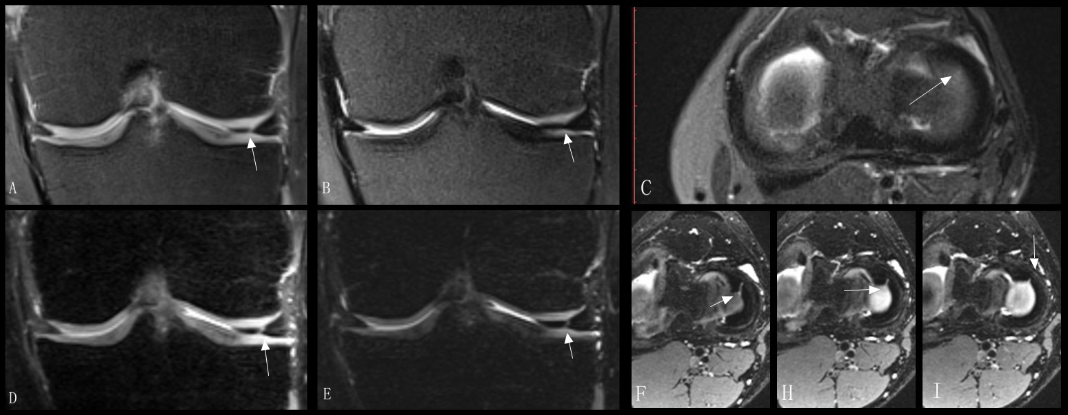
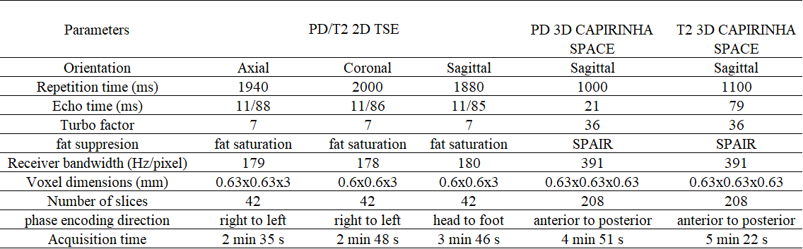
This study was approved by our institutional review board. Twenty patients with varying degrees of knee injury were included in this study (13 men, 7 women; mean age 36.55 years; age range, 17-68 years). Two scan protocols were performed on 3T MRI system (MAGNETOM Skyra, Siemens Healthcare, Erlangen, Germany). Protocol 1: The conventional 2D protocol which used dual echo time in one sequence resulted in a combination of proton density weighted (PDw) and T2 weighted (T2w) turbo spin echo (TSE) contrast. The 2D protocol was applied in transversal, sagittal and coronal imaging planes. Protocol 2: A 3D protocol included two 3D CAIPI-SPACE prototype sequences with both PDw and T2w images. The main parameters of the two protocols are showing in Table 1. Two readers scored the knee joint structure lesions showed on the two protocols using modified MOAKS3. The scoring items included the meniscus morphology, cartilage, bone marrow edema, ligament and effusion. For meniscus morphology, our score method was originated from Lotysch’s4. There were 0-3 scores for each subregion except for ligaments which is graded by the present and absent of tear. Weighted kappa was performed for consistency of the two sequences.Results
The overall presentation of the 20 subjects was shown on table 2. For meniscus tear, a total of 18 subregions in 9 patients were scored for grade 3 in protocol 2, while only 14 of them were detected by protocol 1 (score of 1 or 2). The consistency between the two protocols was substantial for meniscus morphology changes (k=0.66). What’s more, 3D CAIPI-SPACE can detect more cartilage lesions than the conventional 2D MR (17 subregions vs 9 subregions) and can better depict the full-thickness loss of cartilage (8 subregions vs 2 subregions). The consistency was substantial for patella cartilage (k=0.79) and was moderate for femur cartilage (k=0.50). For bone marrow edema, there was substantial to almost perfect consistency between the two protocols (k=0.79, 0.82, 0.85 for tibia, patella and femur respectively). And for anterior cruciate ligament tear and effusions, there was no difference between the two protocols.Discussion
3D CAIPI-SPACE enables better depiction of cartilage lesions and meniscus morphology changes attributed to its thin slice and high image quality. It is easier for 3D SPACE sequence to distinguish cartilage edges from effusions compared with conventional 2D protocol as the 3D SPACE shows a better image contrast between the cartilage and the effusions. What’s more, the isotropic volumetric imaging of 3D SPACE allows for multi-planar reconstruction which enables to provide more diagnostic information of structures, such as the different oriented directions of different ligaments and tendons. Therefore, the diagnostic performance of 3D CAIPI-SPACE in detecting bone marrow lesions was not inferior to the conventional 2D knee MRI protocol. Furthermore, the total acquisition time for SPACE protocol was about ten minutes, which was similar to the conventional knee MRI protocol with better image quality and more clinical information. However, its diagnostic performance needs to be further studied with correlation to arthroscopy and should be applied in more patients with different kinds of diseases.Conclusion
3D CAIPI-SPACE sequence may be expected to replace the conventional 2D clinical protocol in the examination of patient with knee injuries and may be able to provide more morphological information than the conventional 2D protocol.Acknowledgements
No acknowledgement found.References
1. Kalia V, Fritz B, Johnson R, et al. CAIPIRINHA accelerated SPACE enables 10-min isotropic 3D TSE MRI of the ankle for optimized visualization of curved and oblique ligaments and tendons. European radiology 2017;27(9):3652-61. doi: 10.1007/s00330-017-4734-y [published Online First: 2017/01/25]
2. Fritz J, Fritz B, Thawait GG, et al. Three-Dimensional CAIPIRINHA SPACE TSE for 5-Minute High-Resolution MRI of the Knee. Investigative radiology 2016;51(10):609-17. doi: 10.1097/rli.0000000000000287 [published Online First: 2016/05/18]
3. Hunter DJ, Guermazi A, Lo GH, et al. Evolution of semi-quantitative whole joint assessment of knee OA: MOAKS (MRI Osteoarthritis Knee Score). Osteoarthritis and cartilage 2011;19(8):990-1002. doi: 10.1016/j.joca.2011.05.004 [published Online First: 2011/06/08]
4. Crues JV, 3rd, Mink J, Levy TL, et al. Meniscal tears of the knee: accuracy of MR imaging. Radiology 1987;164(2):445-8. doi: 10.1148/radiology.164.2.3602385 [published Online First: 1987/08/01]
Figures