1401
The value of 30°flexed knee MRI in evaluating anterior cruciate ligament tears1Radiology, Peking University Third Hospital, Beijing, China
Synopsis
The study aimed to investigate the value of 30°flexed knee MRI in evaluating anterior cruciate ligament (ACL) tears. 64 patients with knee trauma and hospitalized for arthroscopy were included and performed knee MRI in slightly flexed(about 17°) and 30° flexed positions successively. Images of both positions have high sensitivity, specificity and accuracy in diagnosing ACL tears.30°flexion images were superior to 17°in delineating ACL full length, torn ACL’s disrupted sites and ligament remnants, while no significant difference was found in delineating ACL double-bundle structure. Thus 30°flexed knee MRI was recommended in patients suspecting ACL tears.
Introduction
Anterior cruciate ligament(ACL) is an important structure to maintain the stability of the knee joint. Magnetic resonance imaging(MRI) has become the preferred imaging diagnosis method for ACL and other combined injuries.Current knee MRI coils are roughly divided into two categories: the flexible surface coil,in which knees are scanned in extension;the standard knee coil, in which knees are actually scanned slightly flexed(about 17 °)1.As previously reported2-6,MRI scans in knee flexion(various angles) is superior to extension in delineating ACL and its tears(especially in the femoral attachment). However, few studies have reported the differences between flexed and slightly flexed MRI knee scan in describing and diagnosing ACL tears.Our research aims to investigate the value of 30° flexed knee MRI in evaluating anterior cruciate ligament (ACL) tears compared with 17° flexed knee MRI.Methods
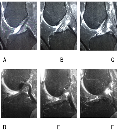
From January 2018 to July 2018,64 patients with a clear history of knee trauma and hospitalized for knee arthroscopy were included and performed 3.0T knee MRI(United Imaging 770,Shanghai,China) in slightly flexed(about 17°) and 30° flexed positions successively,all using standard FSE protocol.Total scan time was 22min(11min for the 30° flexed position).Subsequent arthroscopic examinations in all cases confirmed correct diagnosis in 25 intact ACLs and 39 completely torn ACLs.
The 17° and 30° flexed knee MRI images were reviewed by two experienced radiologists.Each radiologist diagnosed separately whether the ACL was intact,partly torn or completely torn. The delineation of ACL full length, double-bundle structure and torn ACL’s disrupted sites combined with ligament remnants were rated separately.
Wilcoxon test was used to analysis the difference in ratings of ACL full length,double-bundle structure and torn ACL’s disrupted sites combined with ligament remnants. With arthroscopy result as the gold standard, sensitivity, specificity and accuracy of both images in the diagnosis of ACL rupture were calculated.Kappa coefficient analysis were used to evaluate the consistency of the accuracy of diagnosis.The difference was considered significant at P<0.05.
Results
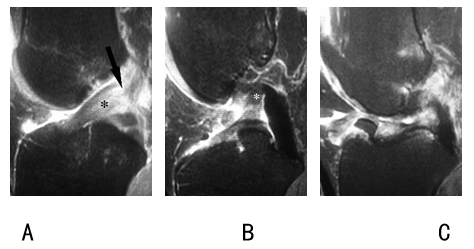
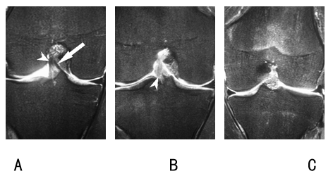
The diagnosing sensitivity, specificity and accuracy of ACL tears were respectively 97%/100%,100%/100% and 98%/100% in slightly flexed and 30°flexed knee MRI, with good consistency between the two positions(Kappa value=0.97). 30°flexion images were rated superior to slightly flexion images in delineation of ACL full length and torn ACL’s disrupted sites combined with ligament remnants(both P<0.05),while no statistical difference was found in rating the delineation of ACL double-bundle structure(P=0.223).Discussion
In this study, we selected 30 ° flexed position to compare with slightly flexed position (about 17 °), because flexion more than 30 ° knee scan may have disadvantages in showing other knee injuries,such as patellofemoral joint stability7.Results show that both two positions have high accuracy,sensitivity and specificityin the diagnosis of ACL tears good consistency.Moreover, 30° flexed position is superior to slightly flexed postion in delineation of ACL full length and torn ACL’s disrupted sites combined with ligament remnants.This might because the anterior edge of the ACL is blurred due to lack of separation between ACL femoral attachment and the intercondylar area at small angle flexion. Thus 30° flexed knee MRI scan could better display ACL tears,for ACL femoral attachment is frequently involved in ACL tears. ACL is thought to consist of two bundles of fibers:anteromedial bundle(AMB) and posterolateral bundle(PLB).When the knee extends,AMB relaxes and PLB tightens.With the flexion of the knee joint, AMB tightens and PLB relaxes, the two fiber bundles twists8.Our study found that 30 ° flexed MRI images displayed ACL double bundle structure not better than slightly flexed position.It is speculated that because the angle changes little in two positions, the bundles are not well-twisted.As far as the authors know, this study is the first to explore the effect of flexed knee MRI scan on the display of ACL double-bundle structure.Conclusion
Both slightly flexed and 30°flexed knee MRI have high sensitivity, specificity and accuracy in diagnosing ACL tears. 30°flexion images were superior to slightly flexion images in delineation of ACL and its tears, while no significant difference was found in delineation of ACL double-bundle structure. Thus 30°flexed knee MRI was recommended in patients with suspected ACL tears.Acknowledgements
No acknowledgement found.References
1.Niitsu M, Ikeda K, Itai Y. Slightly flexed knee position within a standard knee coil: MR delineation of the anterior cruciate ligament[J]. Eur Radiol,1998,8(1):113-115.
2.Niitsu M, Ikeda K, Fukubayashi T, et al. Knee extension and flexion: MR delineation of normal and torn anterior cruciate ligaments[J]. J Comput Assist Tomogr,1996,20(2):322-327.
3.Pereira E R, Ryu K N, Ahn J M, et al. Evaluation of the anterior cruciate ligament of the knee: comparison between partial flexion true sagittal and extension sagittal oblique positions during MR imaging[J]. Clin Radiol,1998,53(8):574-578.
4.Muhle C, Ahn J M, Dieke C. Diagnosis of ACL and meniscal injuries: MR imaging of knee flexion versus extension compared to arthroscopy[J]. Springerplus,2013,2(1):213.
5.Guenoun D, Vaccaro J, Le Corroller T, et al. A dynamic study of the anterior cruciate ligament of the knee using an open MRI[J]. Surgical and Radiologic Anatomy,2017,39(3):307-314.
6.Nenezic D, Kocijancic I. The value of the sagittal-oblique MRI technique for injuries of the anterior cruciate ligament in the knee[J]. Radiol Oncol,2013,47(1):19-25.
7.Becher C, Fleischer B, Rase M, et al. Effects of upright weight bearing and the knee flexion angle on patellofemoral indices using magnetic resonance imaging in patients with patellofemoral instability[J]. Knee Surg Sports Traumatol Arthrosc,2015,25(8):2405-2413.
8.Ng A W H, Lee R K L, Ho E P Y, et al. Anterior cruciate ligament bundle measurement by MRI[J]. Skeletal Radiology,2013,42(11):1549-1554.
Figures