1394
High-resolution ultra-short echo-time (UTE) ex-vivo imaging of Achilles tendons at 9.4T by water content alteration1Medical Physics Group, Institute of Diagnostic and Interventional Radiology, Jena University Hospital - Friedrich Schiller University Jena, Jena, Germany, 2Julius Wolff Institute and Center for Musculoskeletal Surgery, Charité – Universitätsmedizin Berlin, Berlin, Germany, 3Zuse Institute Berlin, Berlin, Germany, 4Michael Stifel Center for Data-driven and Simulation Science Jena, Friedrich Schiller University Jena, Jena, Germany, 5Abbe School of Photonics, Friedrich Schiller University Jena, Jena, Germany, 6Center of Medical Optics and Photonics, Friedrich Schiller University Jena, Jena, Germany
Synopsis
To enable high-resolution imaging of ex-vivo ovine and porcine Achilles tendons, samples were extracted and stored in distilled water for up to 7 days to induce water intake and prolonged T2. One sample was measured continuously to quantify the change in T2 over time, while the other/second sample was used to acquire high-resolution structural images using a cryogenic measurement coil at 9.4T.
Introduction
Ex-vivo imaging of tendons at high field strengths is challenging due to very short T2 relaxation times. Typically, ultra-short echo-time (UTE) acquisition techniques1 are used; nonetheless, these methods have disadvantages, including T2-blurring2, long acquisition times when using short non-selective RF pulses, or an increased sensitivity against eddy current induced errors in the slice profile3 when applying 2D half pulses. Increasing T2 by purposefully orienting the sample along the magic angle of 54.7° with respect to B0 is also challenging due to the limited bore and coil diameters, specifically in case of high field, small-bore scanners. This issue is even aggravated when it comes to achieving very high spatial resolution by utilizing even smaller cryogenic coils. In the current work, we present a method to increase T2 of ex-vivo tendon samples by altering the water content. The change of T2* over time was quantified and ultra-high-resolution structural imaging was performed.Methods
To perform ex-vivo imaging of tendons, samples are typically embedded in physiological saline solution to avoid drying. The use of isotonic solutions furthermore balances effectively osmolar concentrations and avoids tissue swelling or shrinking. Placing a sample in a non-isotonic solution, such as distilled water, a deliberate water intake can be induced.
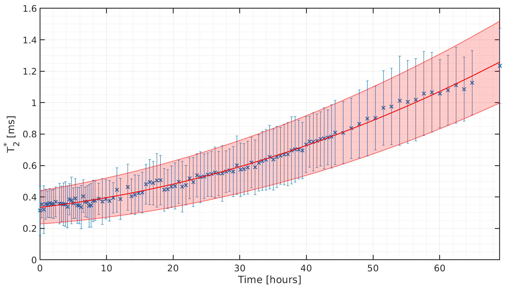
To test the influence of water intake on T2*, a fresh ovine Achilles tendon without any signs of pathology was placed in a tube filled with distilled water and measured continuously for 69 hours. Measurements were performed on a 9.4T Bruker BioSpec-USR 94/20 MR scanner using a 35mm volume coil and gradient insert (BGF6S-100 for BGA12S2). Since the vendor-supplied 2D UTE imaging sequence only supports single echoes, multiples of this sequence were interleaved using manual echo train shifting with echo times of 0.35ms, 0.40ms, 0.50ms, 0.80ms, 1.50ms, 3.00ms, 5.00ms, 8.00ms and 11.00ms. Each echo acquisition used the following parameters: 164x164 acquisition matrix; 3.3x3.3 cm² FoV; 0.8mm slice thickness, 15ms TR; 1 min TA. This set of echo train-shifted acquisitions was repeated 113 times, spanning a total of 1017 acquisitions over 69 hours. The acquired images were combined into a single dataset before performing squared exponential fitting over the echo direction in each voxel. Mean T2* values averaged over a manually outlined region-of-interest within the tendon were extracted for each time point.
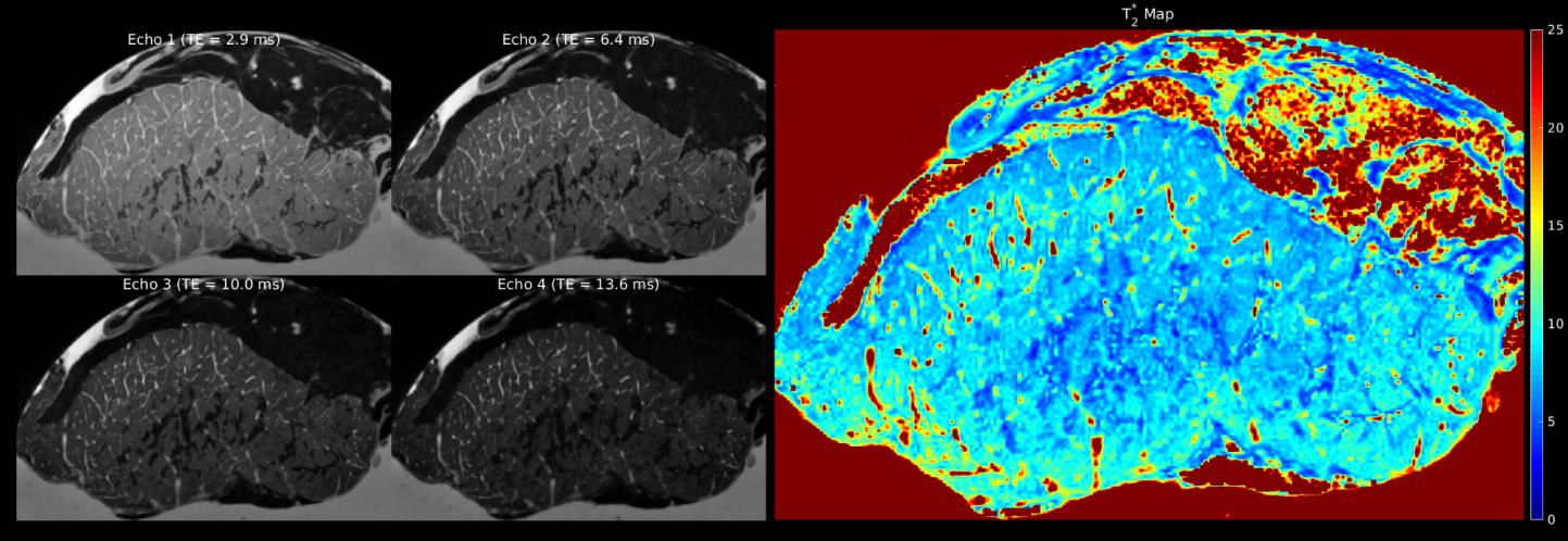
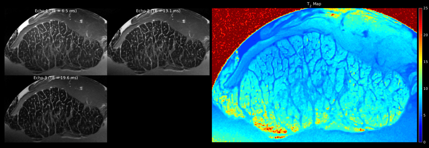
For high-resolution structural imaging, a fresh porcine Achilles tendon without any signs of pathology was placed in a tube filled with distilled water for 7 days before being measured using the same 9.4T scanner. In this experiment, a two-channel quadrature cryoprobe was used instead of the gradient insert. Both 2D multi-echo gradient-echo (MGE) and multi-echo spin-echo (MSE) sequences were used with the following parameters: 192x308 acquisition matrix; 8x13 cm² FoV; 40x40 µm² spatial in-plane resolution. The MGE acquisition used the following parameters: TR 4000 ms; 3 averages; 70 slices with thickness of 200 µm; 4 echoes with TEs of 2.9ms, 6.4ms, 10.0ms and 13.6ms, TA 1 h. The MSE used the following parameters: TR 8000ms; 3 echoes with TEs of 6.5ms, 13.1ms and 19.6ms; TA 1h 22min; and otherwise matched parameters. All images were acquired with fat saturation.
Results
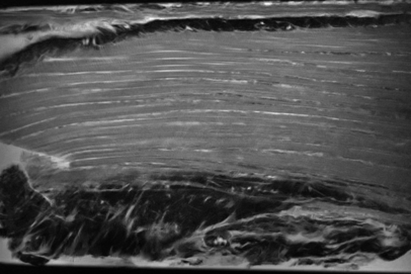
Over the 69-hour measurement, T2* in the ovine Achilles tendon increased from 0.32ms to 1.24ms, as shown in Figure 1. An increase in the volume of the tendon was not observed on the 2D UTE images. The increase in T2* allowed to acquire high-resolution gradient (Fig. 2) and spin echo (Fig. 3) images of the porcine Achilles tendon. Following one week in distilled water, the corresponding mean T2* and T2 values were 7.7ms and 8.8ms, respectively. The directional structure of the collagen fiber bundles could be visualized clearly on coronal images (Fig. 4).Discussion and Conclusion
By purposefully storing ex-vivo ovine and porcine Achilles tendon samples in distilled water, the T2/T2* relaxation times increased significantly, which enabled application of non-UTE imaging sequences. Although the induced water intake influences quantitative tissue analysis and should thus be correspondingly considered, this approach enables high-resolution structural imaging, including spin-echo-based imaging sequences, which cannot be achieved with UTE techniques. The duration of the storage of the tendon in water depends on the desired imaging application for optimizing parameters, such as sample degradation versus increases of T2. From our experiments, no volume changes within the tendon could be observed over the duration of 69 hours. One explanation for not observing significant sample swelling could be the molecular structure of tendons which by itself can bind water by forming a hydration sheet4. Measureable sample swelling could also occur for even longer duration of water intake.Acknowledgements
No acknowledgement found.References
1. Herrmann KH, Krämer M, Reichenbach JR. Time Efficient 3D Radial UTE Sampling with Fully Automatic Delay Compensation on a Clinical 3T MR Scanner. PLoS One. 2016;Mar14;11(3):e0150371
2. Rahmer J, Börnert P, Groen J, Bos C. Three-dimensional radial ultrashort echo-time imaging with T2 adapted sampling. Magn Reson Med. 2006;55(5):1075-1082
3. Josan S, Pauly JM, Daniel BL, Pauly KB. Double Half RF Pulses for Reduced Sensitivity to Eddy Currents in UTE Imaging. Magn Reson Med. 2009;61;1083-1089
4. Fullerton GD, Rahal A. Collagen Structure: The Molecular Source of the Tendon Magic Angle Effect. 2007;25;345-361
Figures