1385
Minimized PET Attenuation in PET-MRI Knee Scanning with Flexible, Screen-Printed MR Coils1Radiology, Stanford University, Stanford, CA, United States, 2Department of Radiology and Biomedical Imaging, University of California - San Francisco, San Francisco, CA, United States
Synopsis
Simultaneous PET-MRI imaging is an exciting new technology for quantitative assessment of whole-joint disease in osteoarthritis (OA). Attenuation correction (AC) for MRI hardware is a challenge for PET quantification in hybrid PET-MRI systems. In this work, we tested a new, lightweight, screen-printed, flexible, 12-channel phased-array MR coil which reduces PET photon attenuation from MR hardware in a PET phantom to below 3%, potentially removing the need to correct for it. Further, the close proximity of coil elements to the knee show promise for high SNR MRI knee imaging.
Introduction
Simultaneous PET-MRI imaging is an exciting new technology for quantitative assessment of whole-joint disease in osteoarthritis (OA)1. In particular, PET adds an opportunity to add important functional information regarding bone metabolism to the widely utilized MRI methods for evaluation of soft-tissue morphology and microstructure. However, MR hardware used with hybrid PET-MRI systems may affect both qualitative and quantitative accuracy of PET images2. While rigid, stationary MR hardware are commonly corrected for in MR-based attenuation correction (MRAC), flexible knee coils are currently disregarded as their position and individual geometry is challenging to account for. In this work, we evaluate the potential of new, flexible, screen-printed MRI coils to minimize the PET photon attenuation in PET-MRI knee scanning.Materials and Methods
Imaging experiments were performed on a 3T PET-MRI hybrid system (GE Healthcare, Milwaukee, WI) using: (1) a screen-printed 12-channel receive-only coil3 (Inkspace Imaging, Berkeley, CA), (2) a 16-channel flex receive-only coil (NeoCoil, Pewaukee, WI), and (3) a 18-channel transmit/receive knee coil (QED Electronics, Cleveland, OH). Phantom experiments were performed using a uniform cylindrical germanium-68 [68Ge] phantom. PET imaging of the phantom was performed for 5 minutes with and without the various MR coils, repeated multiple times with repositioning. Non-attenuated-corrected (NAC) PET activity maps were reconstructed for each acquisition. Lastly, one subject was scanned under an approved university IRB to evaluate initial image quality for knee scanning with the screen-printed coil.Results and Discussion
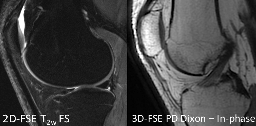
Figure 1 shows axial PET activity maps through a central slice with no coil present as well as with the presence of each coil tested. The screen-printed coil showed the smallest decrease in activity compared with the 16-ch flex coil and the latest state-of-the-art T/R knee coil. This attenuation was seen to be fairly consistent across the length of the PET bed (26 cm) for the screen-printed coil, but was more concentrated around the center of the field for the other coils (where the majority of largely attenuating coil elements are located) [Figure 2]. The screen-printed coil had a mean attenuation of 2.9 ± 0.7% with a local slice maximum of 4.1%. This was an improvement over the currently utilized 16-ch flex coils which are also relatively PET transparent compared to standard knee coils [Table 1]. At 3 percent attenuation, these screen-printed coils offer improved PET SNR and may remove the need for MRAC of MRI hardware. Further, the close proximity of coil elements to the knee show promise for high SNR MRI knee imaging [Figure 3]. Further work is necessary to fully evaluate the MRI performance of these coils as well as their attenuation properties in an in vivo setting.Conclusion
We have shown the feasibility of minimized PET attenuation, with high-SNR MR knee imaging using screen-printed flexible coils.Acknowledgements
This work was funded by GE Healthcare, the William K. Bowes Jr. Stanford Graduate Fellowship, and National Institute of Health (NIH) grants K99EB022634, R01EB002524, K24AR062068 and R01CA212148References
1. Kogan F, Broski S, Yoon D, Gold G. Applications of PET-MRI in Musculoskeletal Disease. Journal of Magnetic Resonance Imaging. 2018; 48(1): 27-47.
2. Wagenknecht G, Kaiser HJ, Mottaghy FM, Herzog H. MRI for attenuation correction in PET: methods and challenges. Magma 2013;26(1):99-113.
3. Corea JR, Flynn AM, Lechene B, Scott G, Reed GD, Shin PJ, Lustig M, Arias AC. Screen-printed flexible MRI receive coils. Nat Commun 2016;7:10839.
Figures