1380
Diagnostic value of MR imaging with metal artifact reduction sequences in local recurrence of malignant bone tumor after joint replacement1Radiology, Ruijin Hospital, School of Medicine, Shanghai Jiao Tong University, Shanghai, China
Synopsis
Effective imaging evaluation after joint replacement is important for patients with malignant bone tumor. This study investigated the diagnostic value of MR imaging with metal artifact reduction sequences (MARS) in local recurrence of malignant bone tumor after joint replacement. The sensitivity, specificity, coincidence rate and Kappa value of MARS in the diagnosis of local recurrence were higher than FSE sequences. The ICC was 0.961 between MR images and pathology in measuring recurrent tumor volume. MR imaging with MARS has significant diagnostic value in local recurrence of malignant bone tumor after joint replacement.
Introduction
Tumor resection combined with joint replacement is currently the standard treatment for malignant bone tumors. However, malignant bone tumors have a higher risk of local recurrence, thus long term follow-up and effective imaging evaluation after surgery are especially important. Prostheses cause severe metal artifacts on conventional magnetic resonance (MR) images, while the metal artifact reduction sequences can effectively reduce metal artifacts and improve image quality. This study intended to investigate the diagnostic value of MR imaging with metal artifact reduction sequences (MARS) in local recurrence of malignant bone tumor after joint replacement.Methods
ninety-four patients who were pathologically diagnosed with malignant bone tumor underwent clinical and imaging follow-up after joint replacement. All patients received MR scans with MARS and FSE sequences. The MR imaging findings of local recurrence were analyzed. Pathological or clinical diagnosis was used as the "gold standard" to assess the effectiveness of MR imaging with MARS in diagnosing local recurrence. The longest and the shortest diameters of the recurrent tumor were measured on MR images to calculate the recurrent tumor volume. The sensitivity, specificity, and consistency rate of MARS were compared with FSE sequences in diagnosing local recurrence of malignant bone tumors. The Kappa test was used to assess the consistency of MARS and FSE sequences with pathology in diagnosing recurrence, respectively. ICC evaluated the consistency of MR images with pathology in measuring the volume of recurrent tumor.Results
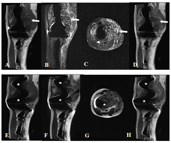
Of the 94 patients, 35 were pathologically or clinically diagnosed as local recurrence after joint replacement. Local recurrence mainly presented as soft tissue masses and bone destruction on MR images. The sensitivity, specificity, coincidence rate and Kappa value of MARS in the diagnosis of local recurrence of malignant bone tumors were 94.3%, 94.9%, 94.7%, and 0.887, which were higher than FSE sequences (sensitivity 80.0%, specificity 84.7%, coincidence rate 83.0%, Kappa value 0.640). The recurrent tumor volume was 58.00 (10.06, 238.00) cm3 measured by MR images, and 49.49 (14.36, 170.38) cm3 by pathological results. The ICC was 0.961 between MR images and pathology in measuring recurrent tumor volume.Discussion
Most patients with malignant bone tumors with local recurrence have a poor prognosis, especially osteosarcoma. Early treatment of local recurrence of tumors can prolong survival, so accurate and early diagnosis of local recurrence is particularly important for prognosis. In this study, we found that the accuracy and the consistency of MARS was higher than FSE sequences in diagnosing local recurrence of malignant bone tumor. MR imaging with MARS has important clinical value in postoperative follow-up for patients with malignant bone tumors. And it may be used for early diagnosis of local recurrence of malignant bone tumors and help to improve the prognosis of patients.Compared with pathological results, the study also confirmed that MR imaging with MARS could assess the volume of local recurrent tumor accurately. The size of recurrent tumor is an independent prognostic factor for osteosarcoma and may influence surgery planning. Recurrent tumor resection is performed when the recurrent lesion is small, while larger recurrent lesions require amputation. Besides, for patients with local recurrence who have received drug treatment, assessing the size of recurrent tumors before and after treatment can help to evaluate the effectiveness.Conclusion
MR imaging with MARS can significantly reduce metal artifacts in scanning area, and diagnose local recurrence. It also has a good consistency with pathology in evaluating the lesion volume, thus it’s recommended for the standard evaluation of postoperative bone tumor.Acknowledgements
This work was financially supported by Action Plan of Major Diseases Prevention and Treatment (2017ZX01001-S12) and Magnetic Resonance (MR)-Dominated Joint Replacement Imaging Evaluation System Research and Clinical Application (17411964900).References
1. He K, Wan Y, Xian S. Risk analysis on infection caused by peripherally inserted central catheter for bone tumor patients[J]. J Can Res Ther 2018;14:90-3.
2. Cipriano C, Griffin AM, Ferguson PC, et al. Developing an Evidence-based Followup Schedule for Bone Sarcomas Based on Local Recurrence and Metastatic Progression[J]. Clin Orthop Relat Res. 2017 Mar;475(3):830-838.
3. National Comprehensive Cancer Network. (NCCN) Clinical Practice Guidelines in Oncology. Bone Cancer, Version 1. 2019. http://www.nccn.org/professionals/physician_gls/pdf/bone.pdf. Accessed August 3, 2018.
4. Ariyanayagam T, Malcolm PN, Toms AP. Advances in Metal Artifact Reduction Techniques for Periprosthetic Soft Tissue Imaging[J]. Semin Musculoskelet Radiol. 2015 Sep;19(4):328-34.
5. Jungmann PM, Agten CA, Pfirrmann CW, et al. Advances in MRI Around Metal[J]. J Magn Reson Imaging. 2017 Oct;46(4):972-991.
6. Molière S, Dillenseger JP, Ehlinger M, et al. Comparative study of fat-suppression techniques for hip arthroplasty MR imaging[J]. Skeletal Radiol. 2017 Sep;46(9):1209-1217.
7. Susa M, Oguro S, Kikuta K, et al. Novel MR imaging method—MAVRIC—for metal artifact suppression after joint replacement in musculoskeletal tumor patients[J]. BMC Musculoskelet Disord. 2015 Dec 4;16:377.
8. Jiang MH, He C, Feng JM, et al. Magnetic resonance imaging parameter optimizations for diagnosis of periprosthetic infection and tumor recurrence in artificial joint replacement patients[J]. Sci Rep. 2016 Nov 14;6:36995.
9. Jungmann PM, Ganter C, Schaeffeler CJ, et al. View-Angle Tilting and Slice-Encoding Metal Artifact Correction for Artifact Reduction in MRI: Experimental Sequence Optimization for Orthopaedic Tumor Endoprostheses and Clinical Application[J]. PLoS One. 2015 Apr 24;10(4):e0124922.
10. Harrison DJ, Geller DS, Gill JD, et al. Current and future therapeutic approaches for osteosarcoma[J]. Expert Rev Anticancer Ther. 2018 Jan;18(1):39-50.
11. Angelini A, Ceci F, Castellucci P, et al. The role of 18F-FDG PET/CT in the detection of osteosarcoma recurrence[J]. Eur J Nucl Med Mol Imaging. 2017 Sep;44(10):1712-1720.
12. Takeuchi A, Lewis VO, Satcher RL, et al. What Are the Factors That Affect Survival and Relapse After Local Recurrence of Osteosarcoma?[J] Clin Orthop Relat Res. 2014 Oct;472(10):3188-95.
13. Grimer RJ, Sommerville S, Warnock D, et al. Management and outcome after local recurrence of osteosarcoma[J]. Eur J Cancer. 2005 Mar;41(4):578-83.
Figures