1374
Differentiation of Soft Tissue Lymphoma from Undifferentiated Sarcoma: ADC Histogram Analysis of Whole Tumor Volume and Single-Slice ADC Measurements at 3T1Radiology, Seoul St. Mary's Hospital, The Catholic University of Korea, Seoul, Korea, Republic of, 2Pathology, Seoul St. Mary's Hospital, The Catholic University of Korea, Seoul, Korea, Republic of, 3Siemens Healthineers, Seoul, Korea, Republic of, 4Orthopedic Surgery, Seoul St. Mary's Hospital, The Catholic University of Korea, Seoul, Korea, Republic of, 5Hematology, Seoul St. Mary's Hospital, The Catholic University of Korea, Seoul, Korea, Republic of
Synopsis
Diffusion-weighted MR imaging should be included in differentiating soft tissue lymphoma from undifferentiated sarcoma to avoid unnecessary surgery in soft tissue lymphoma.
Musculoskeletal Tumors
Purpose: To determine the reliability and accuracy of apparent diffusion coefficient (ADC) histogram analysis of whole tumor volume and single-slice ADC measurements to differentiate soft tissue lymphoma from undifferentiated sarcoma at 3T magnetic resonance (MR) imaging.
Materials and Methods: This retrospective study was approved by the institutional review board and informed consent was waived. Patients with pathologically confirmed soft tissue lymphomas and undifferentiated sarcomas who had undergone 3T MR imaging including diffusion-weighted imaging were included. ADC histogram analysis of whole tumor volume and single-slice ADC measurements were performed using prototype software (Multiparametric Analysis; Siemens Healthcare, Erlangen, Germany). ADC, standard deviation (SD), skewness, and kurtosis were compared between two groups using Mann-Whitney U test. The receiver operating characteristic curve with areas under the curve (AUC) was obtained.
Results: There were 13 patients with soft tissue lymphomas and 12 patients with undifferentiated sarcomas. In ADC histogram analysis, ADC and SD were significantly lower in lymphoma than undifferentiated sarcoma: 650 vs 1278 µm2/sec in ADC; 120 vs 226 in SD (P<.001, P=.017, respectively). Kurtosis of whole tumor volume was significantly higher in lymphoma than undifferentiated sarcoma: 2.0 vs 0.5 (P=.034). On single-slice, ADC and SD were significantly lower in lymphoma than undifferentiated sarcoma: 576 vs 1013 µm2/sec in ADC; 46 vs 96 in SD (P<.001, P=.007, respectively). AUCs of ADC and SD were high in whole volume and single-slice: 0.994 vs 0.968 in ADC and 0.782 vs 0.821 in SD, without significant difference (P=.295, P=.653, respectively). In ADC histogram analysis of whole tumor volume, with cutoff value of 887 µm2/sec, sensitivity and specificity were 92% and 100%. With cutoff value of 143 in SD and 0.51 in kurtosis, sensitivity and specificity were 75% and 77%, respectively. On single-slice ADC measurements, with cutoff value of 641 µm2/sec, sensitivity and specificity were 100% and 85%, while, with cutoff value of SD 37, sensitivity and specificity were 100% and 62%.
Conclusion: Both ADC histogram analysis of whole tumor volume and single-slice ADC measurements may be reliable and accurate in differentiating soft tissue lymphoma from undifferentiated sarcoma.
Acknowledgements
The authors thank Robert Grimm, Siemens Healthcare, Erlangen, Germany for providing the Multiparametric Analysis prototype software.References
1. Kickingereder P, Wiestler B, Sahm F, et al. Primary central nervous system lymphoma and atypical glioblastoma: Multiparametric Differentiation by Using Diffusion-, Perfusion-, and Susceptibility-weighted MR Imaging. Radiology 2014;272:843-850
2. Lee SY, Jee WH, Jung JY, et al. Differentiation of malignant from benign soft tissue tumours: use of additive qualitative and quantitative diffusion-weighted MR imaging to standard MR imaging at 3.0 T. Eur Radiol 2016;26:743-754
3. Politi LS, Forghani R, Godi C, et al. Ocular Adnexal Lymphoma:Diffusion-weighted MR Imaging for Differential Diagnosis and Therapeutic Monitoring. Radiology 2010;256:565-574
Figures
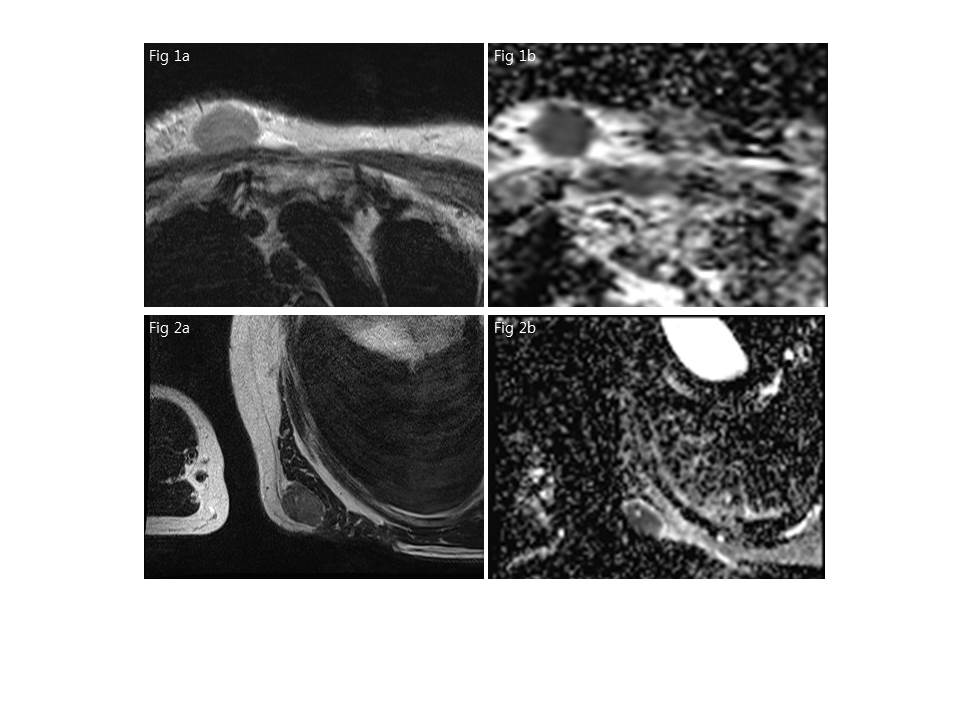
Fig 1. A 89 year-old woman with pathologically confirmed large B cell lymphoma. (a) Axial T2-weighted image (b) On single-slice measurement, ADC 616 µm2/sec and SD 56. On ADC histogram analysis of whole tumor volume, ADC 646 µm2/sec, SD 54, and kurtosis 1.2.
Fig 2. A 65 year-old man with pathologically confirmed undifferentiated sarcoma. (a) Axial T2-weighted image (b) On single-slice measurement, ADC 764 µm2/sec and SD 104. On ADC histogram analysis of whole tumor volume, ADC 885 µm2/sec, SD 292, and kurtosis 1.6.