1366
Cardiovascular Risk Factors for degenerative disc diseases of the thoracolumbar Spine in a Healthy General Population: Results from the KORA MRI Study1Department for Diagnostic and Interventional Radiology, University Hospital Tuebingen, Tuebingen, Germany, 2Department for Trauma and Reconstructive Surgery, BG Trauma Center Tuebingen, Tuebingen, Germany, 3University Hospital Munich, Munich, Germany, 4Department for Diagnostic and Interventional Radiology, Univeristy Hospital Tuebingen, Tuebingen, Germany, 5Radiology, University Hospital Freiburg, Freiburg, Germany
Synopsis
Evidence has grown that disc degeneration may be influenced by cardiovascular risk factors. Thus, the purpose was to assess the association in a healthy sample from the general population. Disc degeneration as well as disc bulging and protrusion of the thoracic and lumbar spine was assessed using the Pfirrmann-score and correlated to risk factors such as obesity, hypertension and diabetes. Hypertension dependently while age and BMI independently associated with disc degeneration. Diabetes increased disc degeneration at Th7/8 and L3/4. Lifestyle conditions had no influence. In conclusion, certain cardiovascular risk factors are associated to disc degeneration and disc bulging and protrusion.
Introduction
Chronic back pain, especially of the lumbar spine with a prevalence above 10%, is a common burden in western nations, and can lead to physical and psychological restrictions1-3. Mostly, no specific cause is found2. However, symptoms can be caused by reduced/imbalanced muscle strength, degeneration of the bony vertebras, reduced intervertebral disc space with disc degeneration (DD), disc bulging (DB) and disc protrusion (DP) as well as a combination of these degenerative changes2-5. Recent evidence has grown, that DD is caused by compromising the serving blood flow of the intervertebral discs due to atherosclerosis and occlusion, which is influenced by cardiovascular risk factors5-7, such as lifestyle conditions (smoking, alcohol, etc.), biochemical/physiological markers (LDL/HDL, triglycerides, blood pressure, diabetes mellitus, obesity, etc.) and subclinical diseases (coronary calcium, etc.)8.
The purpose of this study was to determine the association between demographic and cardiovascular risk factors as well as degenerative intervertebral disk disease in a healthy sample from the general population.
Methods
The cohort of this study was drawn from the KORA Study (Cooperative Health Research in the Augsburg Region) cohort. Healthy volunteers were prospectively enrolled and underwent whole-body magnetic resonance imaging (MRI), including a T2-SS-FSE as well as a dual-echo Dixon sequence of the thorax and abdomen.
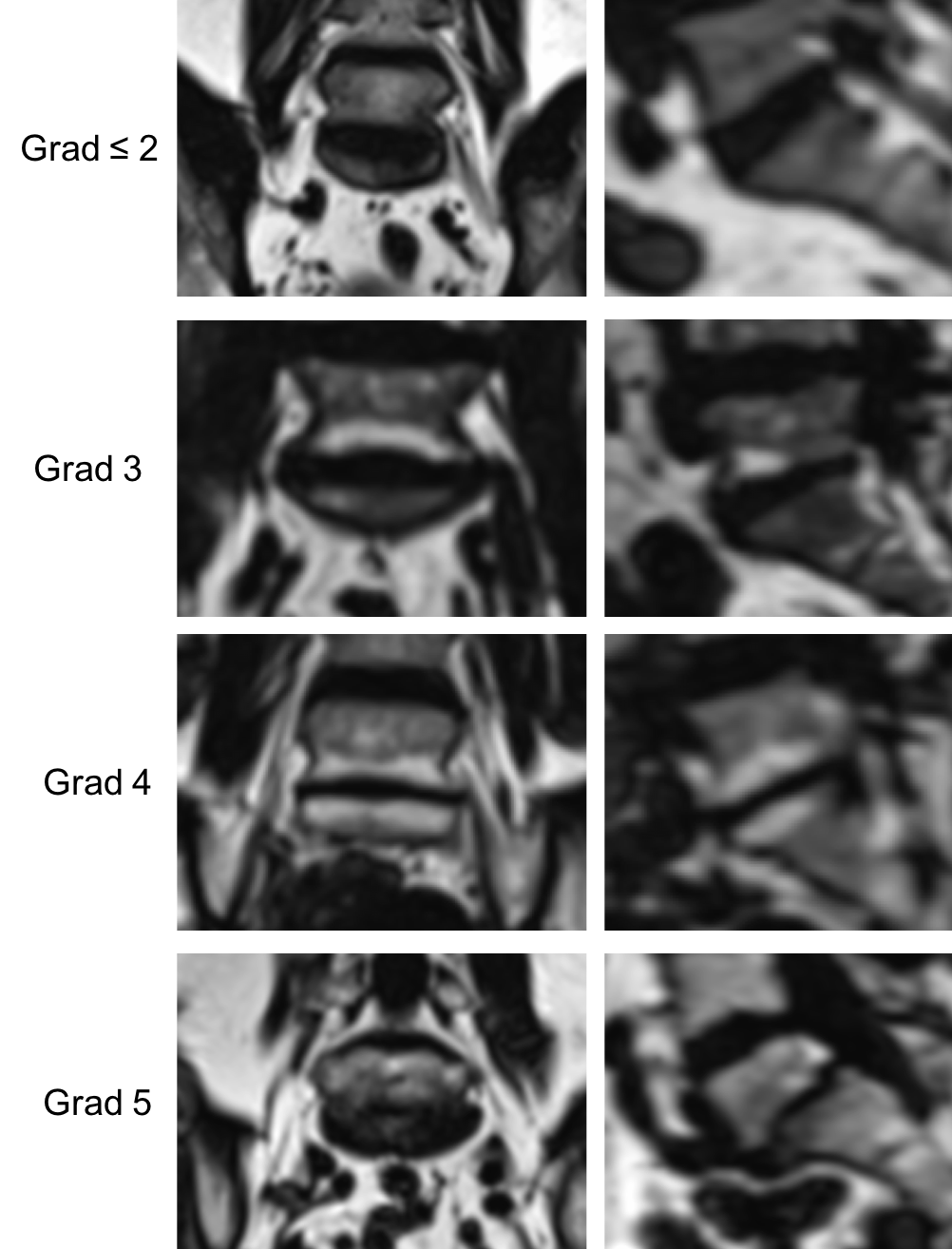
Image evaluation was independently performed in a randomized and blinded fashion to the clinical data by two radiologists with 3 years and 11 years of experience in musculoskeletal imaging. In order to determine intra-reader agreement, 40 cases were again randomized and blinded in order to be reevaluated by the primary reader. DD was assessed with the Pfirrmann-Score, whereby grade 1 is defined as homogenous white structure of the disc, grade 2 as inhomogenous with/without horizontal bands, grade 3 as annulus and nucleus with clear distinction, grade 4 as almost collapsed disc while grade 5 is defined as collapsed disc9 (Figure 1). Aggregation of Grade 1 and 2 as a subgroup was performed since both were classified as low grade DD and a proper differentiation wasn’t distinct in most cases.
Herniation of the discs into the spinal canal were subclassified into DB or DP. Due to anatomical structures, DB was classified as a bulging not exceeding the adjoining vertebrae, while DP was classified as a perimeter of the IVD exceeding the adjoining vertebrae.
Cardiovascular risk factors were obtained as part of the clinical study protocol of KORA.
Results
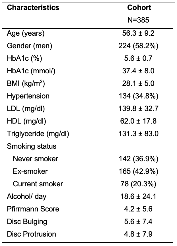
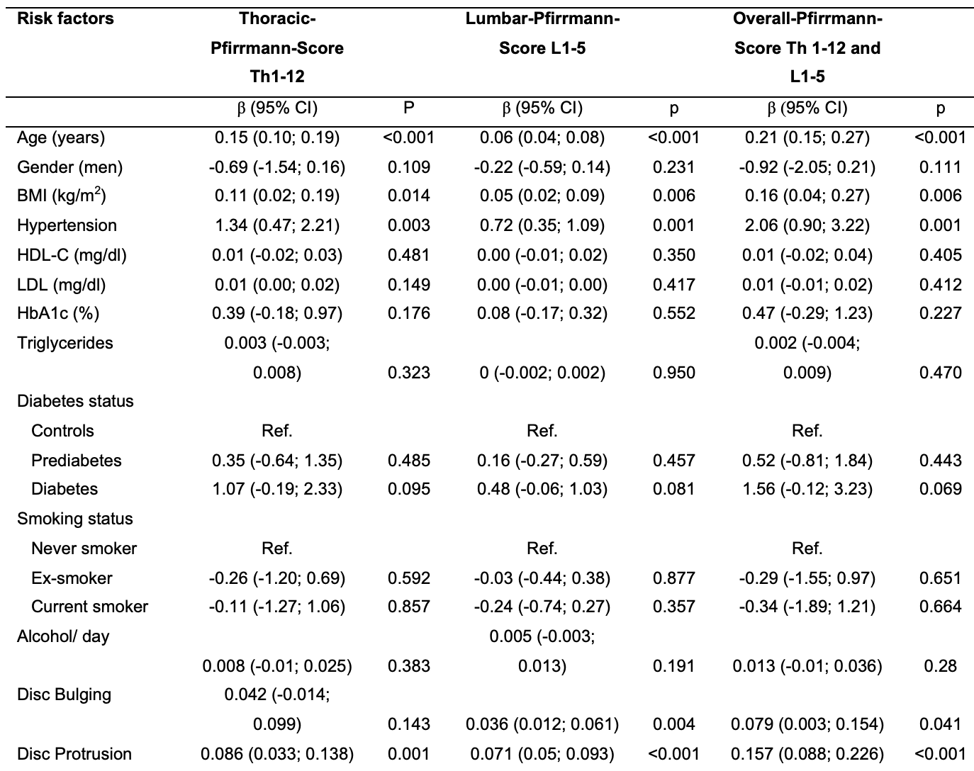
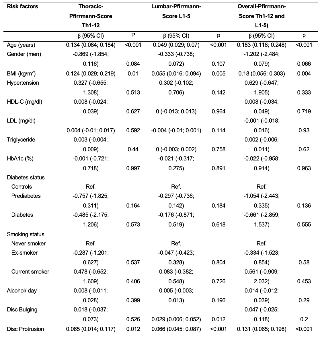
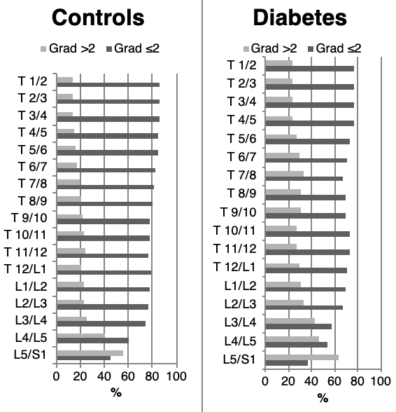
385 of 400 (96.3%) totally enrolled subjects were included (mean age: 56.3 ± 9.2 years, 58.2% males; Figure 2). The overall prevalence of degenerative disc disease was 76.4% (thoracic DD: 40.8%; lumbar DD: 68.1%) whereas disc bulging and protrusion were 37,4% and 27.8%, respectively. Age (p<0.001), BMI (p <0.05) and hypertension (HT) (p <0.05) were associated with DD of the entire spine in univariate analysis, while multivariate analysis only showed an association between age, BMI and DD (p<0.05) (Figure 3 and 4). HT, HDL, LDL, triglyceride (TG), smoking habits and alcohol consumption weren´t significantly associated with DD. Diabetes mellitus (DM) was not association with DB and DP, while significant more degenerative disc changes were found at Th7/8 and L3/4 compared to patients without DM (Figure 5). HT only showed significant association to DB (p<0.05) in multivariate testing.
The intra-readeragreement for evaluation of the thoracic and lumbar intervertebral DD, was almost perfect (85%; Kendall W=0.93) as well as for the inter-reader agreement (87.5%; Kendall W=0.92).Furthermore, DB showed a perfect intra- and inter-reader correlation with 100% accordance (K=1.0), while DP also had a perfect intra-reader convergence (100%; K=1.0) and an almost perfect inter-reader agreement of 95% (K=0,84).
Conclusion
An elevated BMI was associated with an increase of DD and HT was independently associated with DB. DM was associated with increased DD Th7/8 and L3/4, but not with DB and DP.Acknowledgements
No acknowledgement found.References
1. Takatalo J, Karppinen J, Taimela S, Niinimaki J, Laitinen J, Sequeiros RB, et al. Association of abdominal obesity with lumbar disc degeneration--a magnetic resonance imaging study. PLoS One. 2013;8(2):e56244.
2. Abraham I, Killackey-Jones B. Lack of evidence-based research for idiopathic low back pain: the importance of a specific diagnosis. Arch Intern Med. 2002;162(13):1442-4; discussion 7.
3. Steffens D, Maher CG, Pereira LS, Stevens ML, Oliveira VC, Chapple M, et al. Prevention of Low Back Pain: A Systematic Review and Meta-analysis. JAMA Intern Med. 2016;176(2):199-208.
4. Zhang TT, Liu Z, Liu YL, Zhao JJ, Liu DW, Tian QB. Obesity as a Risk Factor for Low Back Pain: A Meta-Analysis. Clin Spine Surg. 2016.
5. Hangai M, Kaneoka K, Kuno S, Hinotsu S, Sakane M, Mamizuka N, et al. Factors associated with lumbar intervertebral disc degeneration in the elderly. Spine J. 2008;8(5):732-40.
6. Shcherbina A, Longacre M. The Association Between Atherosclerosis and Low Back Pain: A Systematic Review. PM R. 2017;9(11):1144-56.
7. Kauppila LI. Atherosclerosis and disc degeneration/low-back pain--a systematic review. Eur J Vasc Endovasc Surg. 2009;37(6):661-70.
8. O'Donnell CJ, Elosua R. [Cardiovascular risk factors. Insights from Framingham Heart Study]. Rev Esp Cardiol. 2008;61(3):299-310.
9. Urrutia J, Besa P, Campos M, Cikutovic P, Cabezon M, Molina M, et al. The Pfirrmann classification of lumbar intervertebral disc degeneration: an independent inter- and intra-observer agreement assessment. Eur Spine J. 2016;25(9):2728-33.
10. Teraguchi M, Yoshimura N, Hashizume H, Yamada H, Oka H, Minamide A, et al. Progression, incidence, and risk factors for intervertebral disc degeneration in a longitudinal population-based cohort: the Wakayama Spine Study. Osteoarthritis Cartilage. 2017;25(7):1122-31.
11. Teraguchi M, Yoshimura N, Hashizume H, Muraki S, Yamada H, Oka H, et al. Metabolic Syndrome Components Are Associated with Intervertebral Disc Degeneration: The Wakayama Spine Study. PLoS One. 2016;11(2):e0147565.
Figures