1356
Evaluation of the risk of osteoporosis in diabetic patients by IDEAL-IQ1Department of Radiology, the First Affiliated Hospital of Dalian Medical University, Dalian Liaoning, China, 2GE Healthcare, Beijing, China
Synopsis
Osteoporosis is characterized by "decreased bone strength and increased risk of fracture”. Osteoporosis caused by diabetes is metabolized caused by decreased bone mass, bone microstructural destruction, increased bone fragility and prone to fracture. It is found that IDEAL-IQ can monitor bone marrow fat changes, diagnose bone marrow lesions or evaluate their functional status.
Purpose
To investigate the correlation between IDEAL-IQ and dual-energy X-ray absorptiometry(DXA)in the assessment of osteoporosis risk in diabetic patients.Materials and Methods
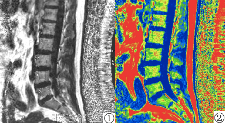
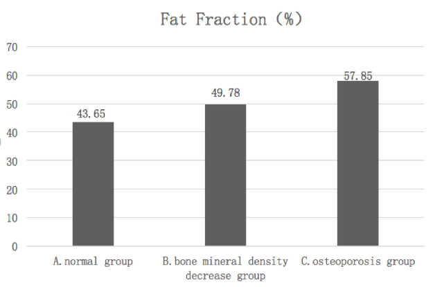
After providing informed consent, 16 diabetic patients underwent magnetic resonance lumbar vertebra IDEAL-IQ sequence scan and dual-energy X-ray absorptiometry (DXA) scan within 24 hours. Volunteers aged 40-70 years, body mass index(BMI)20.21~ 29.74kg/m2. After the dual-energy X-ray absorptiometry (DXA) was performed, all patients with diabetes were divided into three groups: Group A was normal group, group B was bone mineral density decrease group, and group C was osteoporosis group. The IDEAL-IQ sequence automatically generates 6 images, including R2* relaxation rate images, water images, fat images, in-phase and out-phase images. The FF values of vertebral bone marrows were measured on GE AW4.6 post processing workstation(Fig.1). The FF values of lumbar vertebra quantitatively measured by IDEAL-IQ sequence in A, B and C groups were analyzed and compared. The data were analyzed by SPSS 19.0 statistical software, P <0.05 was considered statistically significant.Results
The FF values of lumbar vertebra between group A, B and C were statistically significant(P<0.05), The FF values of group A and group B were significantly smaller than group A and C (P<0.05)(Tab 1).Discussion
Bone marrow is a hematopoietic tissue in the human body. The bone marrow of adults is generally divided into yellow bone marrow and red bone marrow. It is an important hematopoietic tissue and immune tissue, and adipose tissue is an important component of the medullary cavity[1,2]. With the deepening of research, studies have confirmed that bone marrow adipocytes and osteoblasts are derived from the common precursor—bone marrow mesenchymal stem cells[3]. Thus, adipose tissue is one of the important factors affecting the bone marrow microenvironment. Therefore, changes in adipose tissue in the bone marrow will directly affect changes in bone marrow composition, while IDEAL-IQ sequence can monitor changes in bone marrow fat.Conclusion
IDEAL-IQ can quantitatively evaluate the fat fraction of lumbar vertebral and evaluate the risk of osteoporosis in diabetic patients, and it has guiding value for clinical diagnosis and treatment, which is of guiding value for clinical diagnosis and treatment.Acknowledgements
No acknowledgement found.References
[1] Wright NC, Looker AC, Saag KG, et al. The recent prevalence of osteoporosis and low bone mass in the United States based on bone mineral density at the femoral neck or lumbar spine[J]. J Bone Miner Res, 2014,29(11):2520-2526.
[2] Geith T, Schmidt G, Biffar A, et al. Comparison of qualitative and quantitative evaluation of diffusion-weighted MRI and chemical-shift imaging in the differentiation of benign and malignant vertebral body fractures[J]. Ajr American Journal of Roentgenology, 2012, 199(5):1083-1092.
[3] Bermeo S, Gunaratnam K, Duque G. Fat and bone interactions.[J]. Current Osteoporosis Reports, 2014, 12(2):235-242.
Figures