1354
Magnetic resonance (MR) imaging-based fat signal fraction quantification of paraspinal back muscle: Comparison of T1, T2 and gradient-echo m-Dixon method1Department of Radiology, Kyung Hee University Hospital at Gangdong, Seoul, Korea, Republic of
Synopsis
To compare the T1 and T2 m-dixon sequences with GRE mdixon
sequence in FF quantification of paraspinal muscles and to evaluate the
association of all sequences in FF quantification. Fat fraction quantification
of the paraspinal muscles on three m-Dixon MR sequences were different from one
another. But, they have the same tendency and strong correlation.
Introduction
Sarcopenia is defined as loss of muscle function and volume (1). And, it is recognized as an important risk factor for osteoporosis, physical disability and mortality (2). For the diagnosis of sarcopenia, there has been an attempt to measure muscle volume and fat infiltration on MRI and CT (3). Especially, the accurate quantitative measurement of paraspinal muscle fat / has become an important issue. The spin echo 2 point dixon technique is clinically used to detect fat with broad availability and high resolution. And, m-dixon variants have been developed providing more accurate fat quantification. Especially, Gradient echo based dixon method can provide more precise FF by reducing T1 and T2 bias. But, in clinical setting, this method is not commonly included in L-spine MRI (3). To the best of our knowledge, no study has ever evaluated the direct association of T1, T2 and GRE based m-Dixon imaging /for fat quantifications of paraspinal muscles. Thus, the purposes of this study were to compare the T1 and T2 m-dixon sequences with GRE mdixon sequence in FF quantification of paraspinal muscles /and to evaluate the association of all sequences in FF quantification
Methods
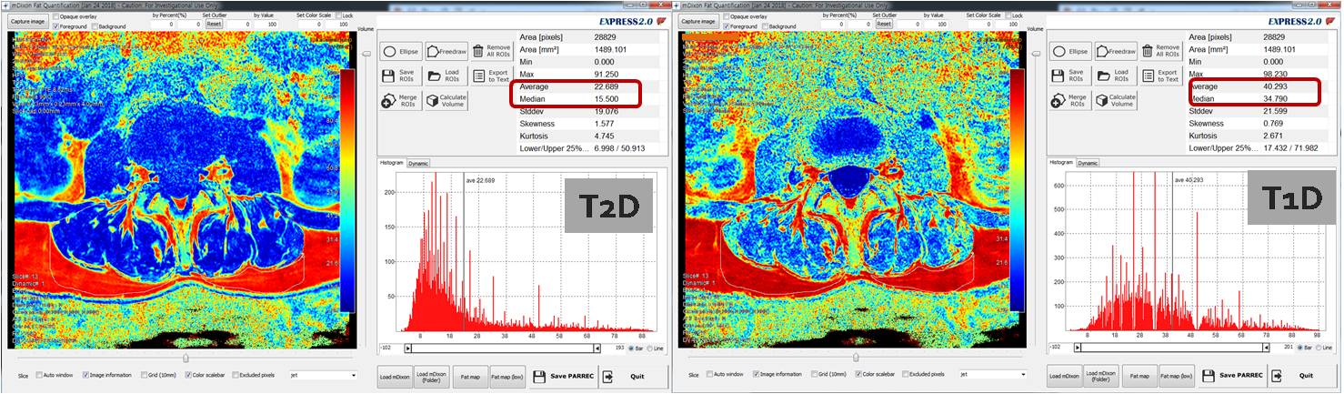
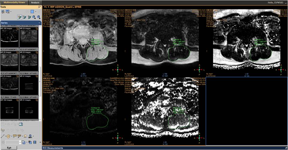
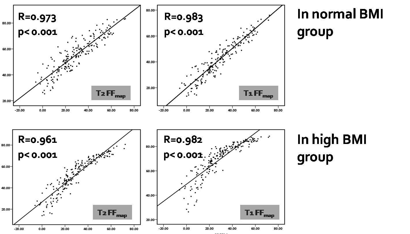
From February 2018 to march 2018, 19 consecutive patients who have taken L spine MRI were recruited. And, subjects were divided into 2 groups, including 11 normal BMI and 8 high BMI subjects. Using 3 tesla MR system, We performed the conventional MRI including T1W and T2 m-dixon sagittal planes. Additionally, T1 and T2 2 point TSE m-Dixon and GRE m-Dixon axial images were acquired. We obtained the ROIs manually on fat only images at 5 disc levels in L-spine. T1 and T2 dixon measurements were performed using a m-dixon quant software. Drawn ROIs were directly copied onto the automatically reconstructed FF map. Statistically, repeated measures ANOVA and pearson’s correlation were performed.Results
In repeated measured ANOVAs, FF quantifications on three sequences are significantly different from one another (p<0.001). In correlation analyses, In both BMI groups, the fat fractions from both sequences showed positive correlation with fat fraction from Quant. The correlation coefficient value higher than 0.9 indicate a very strong linear association (R=0961-0.983).Conclusion
Fat fraction quantification of the paraspinal muscles on three m-Dixon MR sequences were different from one another. But, they have the same tendency and strong correlation. T1 m-Dixon and T2 m-Dixon MR sequences may be used helpful in evaluating the fat fraction of the paraspinal muscles in routine clinical setting.Acknowledgements
NoneReferences
1. Boutin RD, Yao L, Canter RJ, Lenchik L. Sarcopenia: Current Concepts and Imaging Implications. AJR AJR Am J Roentgenol. 2015 Sep;205(3):W255-266.
2. Tyrovolas S, Koyanagi A, Olaya B, Ayuso-Mateos JL, Miret M, Chatterji S, et al. The role of muscle mass and body fat on disability among older adults: A cross-national analysis. Exp Gerontol. 2015 Sep;69:27-35.
3. Reeder SB, Hu HH, Sirlin CB. Proton density fat-fraction: a standardized MR-based biomarker of tissue fat concentration.J Magn Reson Imaging. 2012 Nov;36(5):1011-1014.
Figures