1339
Fatty infiltration of paraspinal muscles is associated with bone mineral density of lumbar spine1The Third Affiliated Hospital of Southern Medical University (Orthopaedic Hospital of Guangdong Province), guangzhou, China, 2Stony Brook Medicine, Stony Brook, NY, United States, NY, NY, United States, 3The Fifth Affiliated Hospital of Sun Yet-sen Univeristy, zhuhai, China
Synopsis
Paraspinal muscle fatty infiltration (FI) is an important factor affecting spinal function. However, there is no previous study investigating the relationship between paraspinal muscle FF and spinal BMD. Our study demonstrated that fat fractions of erector
Objectives Fatty infiltration (FI) in skeletal muscles has gained more attention for the evaluation of muscle function and it can be reduced through exercise. Better understanding of the relationship between muscles FI and BMD can potentially help in the development of interventions benefiting musculoskeletal function; improved musculoskeletal function in terms lead to reduced adverse clinical outcomes such as falls and fractures. It is well known that BMD declines and muscle FI increases in aging. However, the relationship between muscle FI and BMD with age controlled has not been previously reported.In this prospective study, we investigate the relationship between paraspinal muscle fatty infiltration (FI) and bone mineral density (BMD).
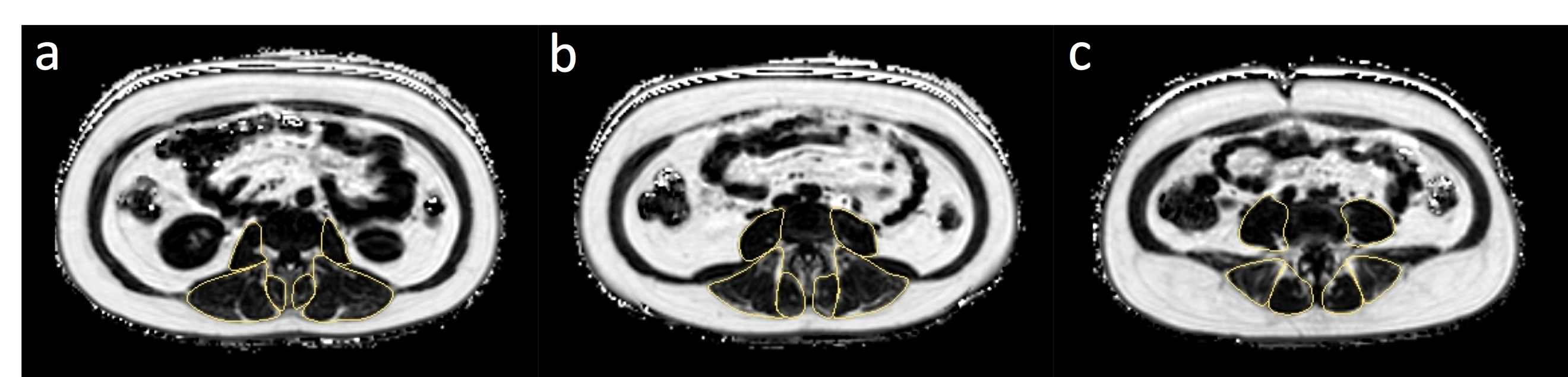
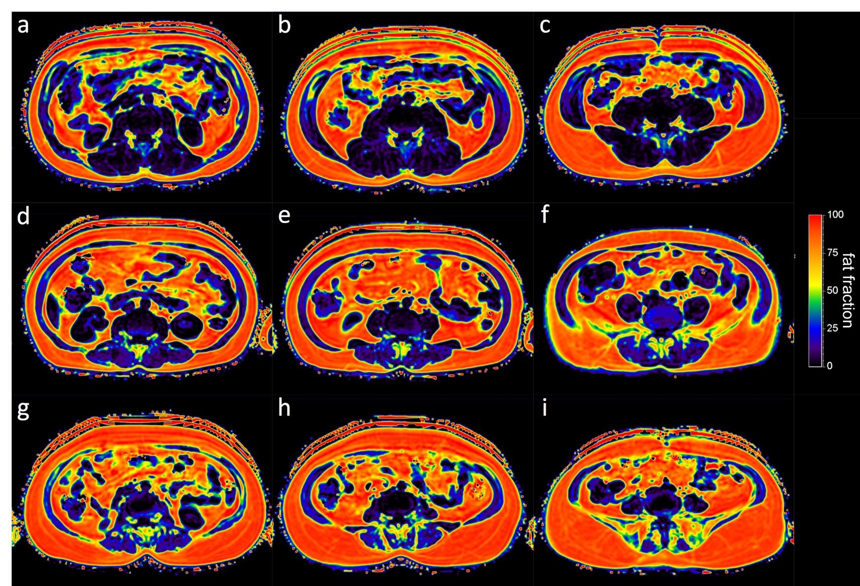
Methods 88 subjects were enrolled in this study (52 females, 36 males; age: 46.6±14.2 years old; BMI: 23.2±3.49 kg/m2). Paraspinal muscle [erector spinae (ES), multifidus (MF) and psoas (PS) ] fat fraction (FF) were measured respectively on axial fat fraction maps at L2/3, L3/4 and L4/5 levels using mDixon Quant. Quantitative Computed Tomography (QCT) was used to access vertebral BMD of L1,L2 and L3.The difference of paraspinal muscle FF among subjects with normal bone density, osteopenia and osteoporosis was tested using one-way ANOVA. The relationship between paraspinal muscle FF and BMD were analyzed by linear regression controlling age, sex and BMI. Partial correlation analysis was performed between paraspinal muscle FF and one variable from age, BMI and sex with the other two controlled.
Results FF of ES, MF and PS of subjects with normal bone density were all significantly less than those with osteopenia and those with osteoporosis (all p<0.001). There is an inverse correlation between paraspinal muscle FF of ES, MF and ES and vertebral BMD after controlling for age, sex and BMI (r: -0.21~-0.29; all p<0.05). The FF of ES and PS had an inverse correlation with BMD after controlling for age and BMI in females but not in males (all p<0.05). There was a moderate correlation between FF of ES,MF,PS and age (r:0.554~0.670, all p<0.001).
Conclusions Our study demonstrated an inverse correlation between paraspinal muscles FI and BMD after adjusting age, sex and BMI. Better understanding the complex relationship between fatty changes of paraspinal muscles and BMD would help us gain more insight into preventing osteoporosis and related complications. Patients with high FI in paraspinal muscles need to strengthen the paraspinal muscles function and reduce the fat. It may decrease bone loss and the risk of osteoporosis fractures. Further prospective multi-center studies should be performed to confirm this hypothesis. In a word, paraspinal muscle FI increased while lumbar BMD decreased after adjusting age, sex and BMD, especially in females.
Acknowledgements
This study has received funding by the National Natural Science Foundation of China (81801653), the Science and Technology Planning Project of Guangdong Province (No.2017B090912006 and No.2014A020211018) and National Institutes of Health R03CA223052. The authors are very grateful for the technical assistance of Shaoyong Hu, Jialing Chen, Xianfu Mo, Wei Fan, Lin Chen and Xiaolong Tan.References
[1].Mengiardi B, Schmid MR, Boos N, et al. Fat content of lumbar paraspinal muscles in patients with chronic low back pain and in asymptomatic volunteers: quantification with MR spectroscopy. Radiology 2006;240(3):786-792. [2].Yoo HJ, Hong SH, Kim DH, et al. Measurement of Fat Content in Vertebral Marrow Using a Modified Dixon Sequence to Differentiate Benign From Malignant Processes. Journal of Magnetic Resonance Imaging 2017;45(5):1534-1544. [3].Beasley LE, Koster A, Newman AB, et al. Inflammation and race and gender differences in computerized tomography-measured adipose depots. Obesity (Silver Spring) 2009;17(5):1062-1069. [4].Rivas DA, McDonald DJ, Rice NP, Haran PH, Dolnikowski GG, Fielding RA. Diminished anabolic signaling response to insulin induced by intramuscular lipid accumulation is associated with inflammation in aging but not obesity. Am J Physiol Regul Integr Comp Physiol 2016;310(7):R561-569. [5].Urrutia J, Besa P, Lobos D, Andia M, Arrieta C, Uribe S. Is a single-level measurement of paraspinal muscle fat infiltration and cross-sectional area representative of the entire lumbar spine? Skeletal Radiol 2018.Figures