1321
Assessment of Cartilage pH Using AcidoCEST-UTE MRI at 3T with Histological Correlation1University of Arizona, Tucson, AZ, United States, 2Department of Radiology, University of California San Diego, San Diego, CA, United States, 3Radiology Service, VA San Diego Healthcare System, San Diego, CA, United States
Synopsis
The poor correlation between structural abnormalities of osteoarthritis (OA) and OA pain complicates treatment and pain management. Acidosis is heavily implicated in pain, and thus may be used to identify areas of pain not associated with structural damage. In this study, we used a pH-sensitive imaging method known as chemical exchange saturation transfer (acidoCEST) MRI to assess acidosis in cadaveric cartilage tissue and assess the relationship between pH and osteochondral vascularity as determined on histology. We show that acidoCEST MRI can measure extracellular pH (pHe) in cartilage, enabling further studies into the complex relationship between acidification, osteochondral channels, and pain.
Introduction
Osteoarthritis (OA) has a considerable impact on the healthcare system. While diagnostic imaging of OA is widely practiced, not all patients presenting with radiographic OA initially report pain.1 The poor correlation between structural abnormalities and pain is problematic; as pain ultimately develops, identification and subsequent treatment of the associated structural anomalies proves difficult.
In OA, two phenomena occur: (1) blood vessels/nerves extend through channels from subchondral bone (SB) into articular cartilage, and (2) damaged cartilage is significantly more acidic.2,3 Acidification is a major component in pain, and even in the absence of structural damage, is sufficient to decrease nociceptor threshold sensitivity and potentiate pain.4,5 A minimally invasive method to measure extracellular pH (pHe) of cartilage could potentially provide a visual map of the chemistry involved in OA pain and have considerable clinical implications.
The purpose of this study was (1) to evaluate the use of chemical exchange saturation transfer (CEST) MRI in assessment of pHe in phantoms and cadaveric cartilage tissue and (2) to assess the relationship between cartilage pHe and osteochondral channels.
Methods
Phantom Preparation:
Liquid phantoms containing iopamidol, iopromide, or iohexol were used since intra-articular injection of these agents is common in the clinic. 0.2M dilutions were prepared using water or phosphate buffered saline (PBS) and pH was adjusted with HCl or NaOH.
Cartilage phantoms containing coins of pure cartilage were created from nine osteochondral cores, harvested from an 80-year-old male donor. Tissues were soaked in 0.4M iopamidol in PBS with various pH ranging from 6.1-7.9 for 96 hours, then placed with soaking solutions in individual syringes. Phantoms were scanned in a container filled with Fomblin.
MRI and Analysis:
Calibration phantoms were imaged with the acidoCEST pulse sequence6 combined with a UTE readout on a 3T clinical scanner (MR750, GE Healthcare, Milwaukee, WI) using an 8-channel transmit/receive knee coil. The 3D UTE-Cones-CEST sequence (Figure 1) parameters are: TR=62ms, TE=0.032ms, Nsp=5, spoke interval τ=5 ms, FA=5°, BW=166 kHz, FOV=12×12×11.2cm3, matrix=160×160×28. CEST contrast was created with a Fermi pulse: duration=32ms, bandwidth=40Hz, two average B1s=5.4/1.1µT, 61 frequency offsets from -1200 to 1200 Hz in 40 Hz increments, with 1min 10s scan time per frequency. Steady-state before acquisition was achieved using an 8s dummy scan.
For the cartilage phantom scan, the sequence parameters were identical to the calibration phantom except for higher resolution (FOV=10×10×12cm3, matrix=160×160×60) and longer scan time (2min 48s per frequency).
AcidoCEST MRI data was analyzed using a ratiometric approach based on distinct RF-powers.7
pHe with Histological Correlation:
pH values on the surface of cartilage were measured at 13 locations with an optical probe (pHOptica, WPI, Sarasota, FL) and visually scored from 0-4 with the International Cartilage Repair Society (ICRS) Classification System. Osteochondral cores were fixed in 10% zinc formalin, decalcified, and cryosectioned, for H&E staining. Vascular density (VD) per mm was determined for channels penetrating the SB, calcified cartilage (CC) and non-calcified cartilage (NCC), as previously described.8 Pearson correlations were performed for pH, ICRS, and VD.
Results and Discussion
pH Phantoms:
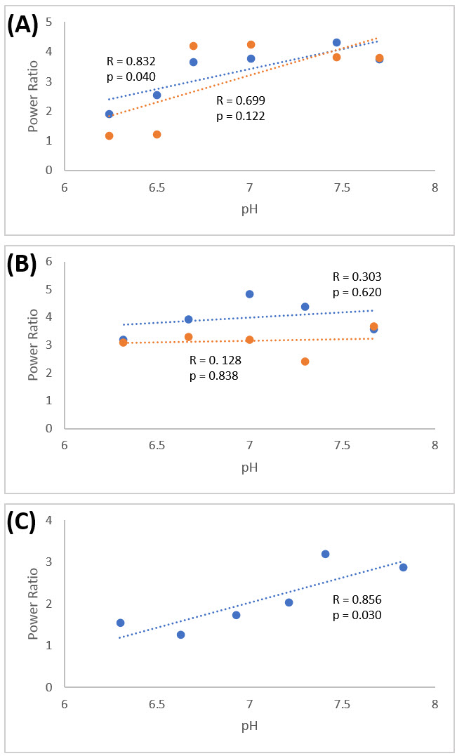
All three contrast agents in solution displayed pH-dependence, with better profiles for iopamidol and iohexol compared with iopromide (Figure 2). Cartilage phantoms with iopamidol demonstrated very strong correlations between power ratio and pH (Figure 3), particularly at 4.2ppm (r=0.983, p=.0004). However, power ratio and pH relationships were slightly different between liquid and cartilage iopamidol phantoms, highlighting the need for calibration using ex vivo tissue phantoms.
Cartilage pHe, Gross and Histology:
No significant correlation was found between pH and ICRS score (p=0.239). A strong significant positive correlation was found between ICRS score and VD terminating in CC (r=0.631, p=0.009), similar to Walsh et al.8, and consistent with their theory that osteochondral angiogenesis/nerve growth may be related to pain in OA. A complex relationship between pHe and VD was observed (Figure 5). A strong negative correlation was found between pHe and VD in SB (r=-0.739, p=0.001). This result may potentially be explained by the strongly acidic environment created by osteoclasts as vascular channels are created in SB. A strong positive correlation was found between pHe and VD in CC (r=0.777, p=0.0004). This result is similar to Walsh et al., who observed higher VD in CC of controls compared with OA patients. A potential explanation may be related to chronic bone loss, whereby released calcium neutralizes acidification.
Conclusion
AcidoCEST-UTE MRI can be used to accurately measure pH in cartilage samples, potentially providing a method to measure cartilage pH in vivo in a manner not previously possible. Complex relationships exist between cartilage pH and osteochondral vascularity, which deserve further study.Acknowledgements
The authors gratefully acknowledge grant support from the VA Rehabilitation R&D Service (I01RX002604), VA Clinical Science R&D Service (I01CX001388) and NIH (R21AR073496 and R01AR062581).References
1. Hannan MT, Felson DT, Pincus T. Analysis of the discordance between radiographic changes and knee pain in osteoarthritis of the knee. J Rheumatol. 2000;27(6):1513-7.
2. Mapp PI, Walsh DA. Mechanisms and targets of angiogenesis and nerve growth in osteoarthritis. Nat Rev Rheumatol. 2012;8(7):390-8.
3. Konttinen YT, Mandelin J, Li TF, et al. Acidic cysteine endoproteinase cathepsin K in the degeneration of the superficial articular hyaline cartilage in osteoarthritis. Arthritis Rheum. 2002;46(4):953-60.
4. Abdelhamid RE and Sluka KA. ASICs mediate pain and inflammation in musculoskeletal diseases. Physiology. 2015 Nov;30(6): 449-59.
5. Reeh PW and Steen KH. Tissue acidosis in nociception and pain. Prog Brain Res. 1996;113:143-51.
6. Jones KM, Randtke EA, Howison CM, Pagel MD. Respiration gating and Bloch-fitting improve pH measurements with acidoCEST MRI in an ovarian orthotopic tumor model. Proc SPIE Int Soc Opt Eng. 2016;27:9788.
7. Wu R, Longo DL, Aime S, Sun PZ. Quantitative description of radiofrequency (RF) power-based ratiometric chemical exchange saturation transfer (CEST) pH imaging. NMR Biomed. 2015 May;28(5):555-65.
8. Walsh DA, McWilliams DF, Turley MJ, et al. Angiogenesis and nerve growth factor at the osteochondral junction in rheumatoid arthritis and osteoarthritis. Rheumatology. 2010 Oct;49(10):1852-61.
Figures