1318
Longitudinal T1ρ mapping of contralateral hip in patients with unilateral cam-type femoroacetabular impingement (FAI)1Medical Imaging, The Ottawa Hospital, Ottawa, ON, Canada, 2Radiology, University of Ottawa, Ottawa, ON, Canada, 3Division of Orthopaedic Surgery, The Ottawa Hospital, Ottawa, ON, Canada, 4Medicine, University of Ottawa, Ottawa, ON, Canada
Synopsis
Cam-type femoroacetabular impingement (FAI) is a major cause of hip osteoarthritis. Quantitative T1ρ MRI has the potential to detect early cartilage degeneration due to its sensitivity to proteoglycan. In this study we performed longitudinally (124 days (average) after surgery and 4.8 years (average) follow up) T1ρ mapping in patients with bilateral (symptomatic and asymptomatic) cam-type FAI on the asymptomatic side after the symptomatic cam-FAI was surgically corrected. The cartilage of the contralateral hip did not show significant proteoglycan depletion and therefore no further degeneration between the initial and the follow up scan was detected. The contralateral hips remained stable.
Introduction
Cam-type femoroacetabular impingement (FAI) is implicated as a mechanism in the premature development of osteoarthritis (OA) of the hip joint1. The aspherical femoral head leads to the eventual development of OA through cartilage damage, especially in the anterolateral region of the weight bearing surface2. Recent research on cam-type FAI using proteoglycan (PG) sensitive imaging techniques (such as T1ρ mapping3) revealed that an early degenerative change is present in the cartilage tissue of symptomatic as well as asymptomatic subjects with cam-FAI4. While symptomatic cam-FAI patients undergo physiotherapy or surgery, longitudinal measurements are necessary to research the asymptomatic cam-FAI in order to develop and monitor treatment strategies. The purpose of this study was to measure the cartilage T1ρ relaxation time in patients with bilateral (symptomatic and asymptomatic) cam-type FAI on the asymptomatic side after the symptomatic cam-FAI was surgically corrected. The subjects were re-imaged longitudinally to determine if any changes occur in the cartilage matrix.Methods
The research ethics board at our institution approved this study and all participants provided informed consent prior to enrollment. 9 patients (8 male, 1 female, mean age: 38.8 years, age range: 27.8 – 48.4 years at day of surgery) undergoing surgical osteochondroplasty for symptomatic cam-type FAI were recruited and enrolled prospectively. Patients were included if they had unilateral painful hip with radiographic evidence of a cam deformity at the femoral head-neck junction in both hips. As part of a larger study protocol, patients underwent computer tomography of both hips and the alpha angle was measured at the anterior (3:00) and anterosuperior (1:30) positions. Subjects underwent T1ρ MRI of the contralateral, asymptomatic hip at an average of 124 days (range: 64 – 285 days) after surgery. A consecutive T1ρ follow-up scan was performed using the same protocol after a mean of 4.8 years (range: 3.2 – 6.0 years). T1ρ mapping was performed on a 1.5T MRI scanner (Siemens) using a spin-lock preparation module combined with a turbo spin echo acquisition scheme in a sagittal oblique orientation. Further parameters: FOV = 180x180mm2, slices = 22, slice thickness = 3mm, matrix = 384x384, resolution = 0.47x0.47mm2, TR = 274ms, TE = 13ms. Five different spin-lock times (TSL) of 12/18/25/35/45ms and a spin-lock field of B1 = 400Hz were used. The total scan time for T1ρ mapping was 21 minutes. The cartilage was segmented as a bilayer in seven to eight slices, starting from the lateral sourcil margin and extending medially. The joint was subdivided into anterior and posterior regions and the slices were divided into lateral, intermediate or medial zones, resulting in six region of interest (ROI) for analysis: Antero-Lateral (AL), Antero-Intermediate (AI), Antero-Medial (AM), Postero-Lateral (PL), Postero-Intermediate (PI) and Postero-Medial (PM) (Figure 1). Statistical analysis (paired t-test) was performed using the statistical toolbox in Matlab, with statistical significance set at p<0.05.Results
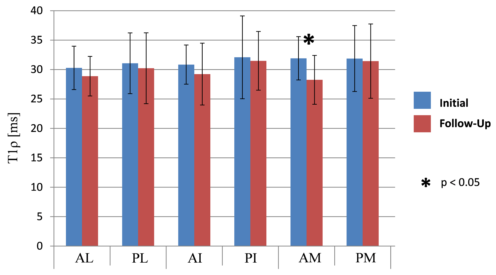
The mean alpha angles were 50° (range: 42 – 64°) at 3:00 and 63° (range: 56 – 70°) at 1:30 position for the surgical side and 54° (range: 50 – 60°) at 3:00 and 63° (range: 51 – 70°) at 1:30 position for the contralateral side. Figure 2 summarizes the results of the hip cartilage T1ρ relaxation times from the six different regions for the initial and the follow-up scan (shown as mean T1ρ values with the standard deviation as error bars). There was no significant difference in T1ρ between two the two scans, except in the anterior-medial (AM) region of the hip. The T1ρ relaxation time decreased there from 31.9±3.7 ms to 28.3±4.2 ms significantly (p< 0.05).Discussion
The findings of this study show that in five of the six analyzed cartilage regions no significant changes in PG content occur within an average of 4.8 years in asymptomatic hips with a cam deformity in subjects who underwent surgical osteochondroplasty on the contralateral side before. In the anterior medial (AM) a small, but significant decrease in T1ρ was detected which implies an increased PG content. The decreased T1ρ relaxation time is possible illustrating a repair/regeneration mechanism within the cartilage in this region. None of the regions showed a significant increase in the T1ρ value implicating that no PG depletion occurred and OA did not proceed in the cartilage tissue. A limitation of the study is the small sample size of only 9 subjects.Conclusion
The cartilage of the contralateral hip in patients with uni-lateral cam-type FAI did not show significant PG depletion based on T1ρ mapping and therefore no further degeneration between the initial and the follow up scan was detected. The contralateral hips remained stable.Acknowledgements
Canadian Institutes of Health Research (CIHR) (Funding number: MOP 97778) for funding of the study.References
[1] Ganz R, Parvizi J, Beck M, et al. Femoroacetabular impingement: a cause for osteoarthritis of the hip. Clin Orthop Relat Res. 2003;(417):112-20.
[2] Beaulé PE, Zaragoza E, Motamedi K, et al. J Orthop Res. 2005 Nov;23(6):1286-92. J Orthop Res. 2005;23:1286-92.
[3] Wheaton AJ, Dodge GR, Elliott DM, et al. Quantification of cartilage biomechanical and biochemical properties via T1rho magnetic resonance imaging. Magn Reson Med. 2005;54:1087-93.
[4] Anwander H, Melkus G, Rakhra KS, et al. T1ρ MRI detects cartilage damage in asymptomatic individuals with a cam deformity. J Orthop Res. 2016;34:1004-9.
Figures