1314
T1ρ at low spin-lock amplitudes is more sensitive to degenerative changes in articular cartilage1Research Unit of Medical Imaging, Physics and Technology, University of Oulu, Oulu, Finland, 2Medical Research Center, University of Oulu and Oulu University Hospital, Oulu, Finland, 3Department of Applied Physics, University of Eastern Finland, Kuopio, Finland, 4Diagnostic Imaging Center, Kuopio University Hospital, Kuopio, Finland, 5Department of Equine Sciences, Utrecht University, Utrecht, Netherlands, 6Department of Orthopaedics, University Medical Center Utrecht, Utrecht, Netherlands, 7School of Information Technology and Electrical Engineering, The University of Queensland, Brisbane, Australia, 8Department of Diagnostics Radiology, Oulu University Hospital, Oulu, Finland
Synopsis
In this study, continuous wave T1ρ scans at various spin-lock amplitudes (γB1 = 100, 200, 300, 400, 500, 600, 800, 1000 and 2000 Hz) were utilized to evaluate multiple articular cartilage regions at increasing distances from a surgically induced lesion in equine specimens. Significant differences were observed between regions adjacent and distant to the lesion, and the differences between the compared sites were larger at lower spin-lock amplitudes. The variations were in agreement with biomechanical properties (determined via indentation testing) of the regions. The findings suggest that T1ρ at low spin-lock amplitudes is more responsive to progressive alterations in articular cartilage.
Introduction
Chondral defects are known to cause gradual degradation in the surrounding tissues as well1,2. Previous studies have evaluated the relationship between T1ρ and progressive deterioration of articular cartilage by comparing degenerated and healthy samples3. The purpose of this study was to investigate the sensitivity of continuous wave T1ρ at varying spin-lock amplitudes to degenerative changes in articular cartilage due to the presence of an adjacent lesion.Methods
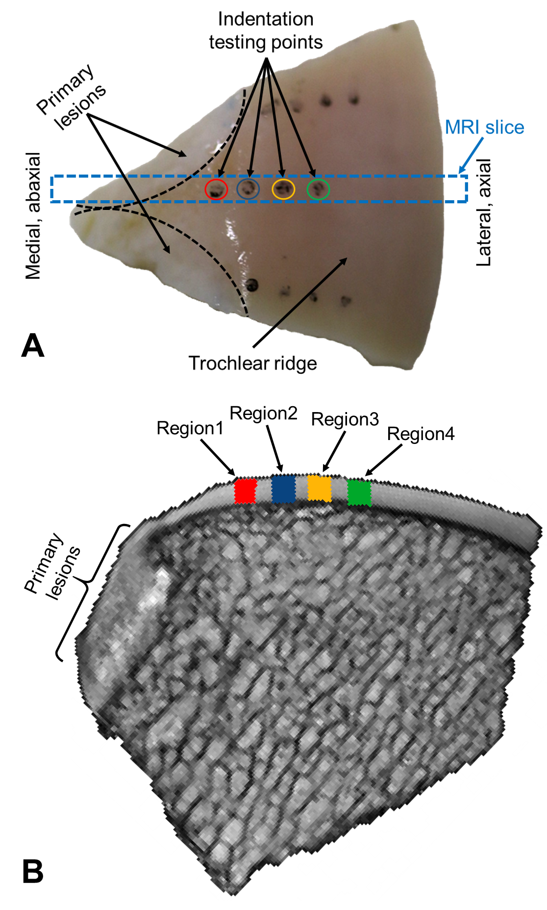
Two circular (d = 10 mm) chondral lesions were created on the medial femoral ridges of both stifle joints of Shetland ponies (aged 4-14 years, N = 7) (Fig. 1A). After 12 months, the ponies were sacrificed and triangular wedge-shaped specimens (25×20×15 mm, n = 13), containing lesions and the surrounding tissue; were obtained from the stifle joints (Fig. 1A). The study had been approved by the local ethical committee of Utrecht University in compliance with the Dutch Act on Animal Experimentation.
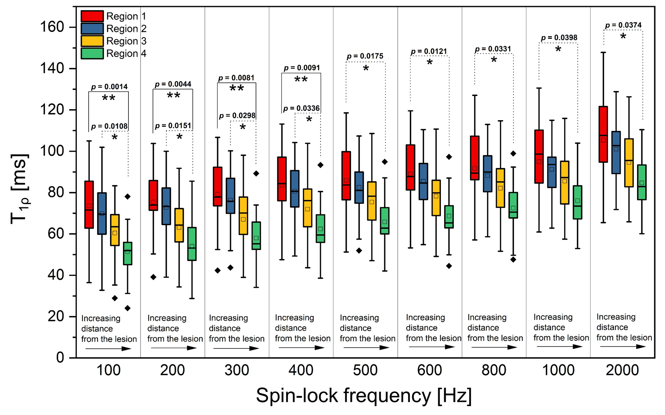
For MR imaging, the specimens were oriented in such way that the articular surface in the imaging region was parallel with the main magnetic field to minimize magic angle related variations between regions. The imaging was performed at 9.4 T using a 19-mm quadrature RF transceiver. A magnetization preparation block, compensating for B1 and B0 field inhomogeneities4, was utilized for T1ρ weighted imaging. T1ρ measurements were carried out with varying spin-lock amplitudes (γB1 = 100, 200, 300, 400, 500, 600, 800, 1000 and 2000 Hz) and seven spin-lock times between 0-128 ms. The preparation was followed by a fast spin-echo readout (TR = 5 s, ETL = 8, TEeff = 4.2 ms, matrix size = 192×192, slice = 1 mm, FOV = 19.2×19.2 mm2).
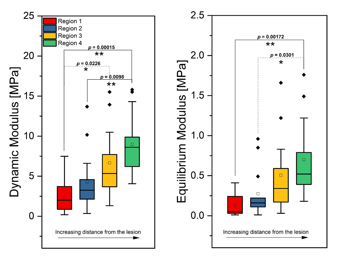
Four full-thickness cartilage regions of interest (ROIs) were defined, matching locations where mechanical properties were measured using indentation testing (Fig. 1B). The indentation protocol consisted of four stress-relaxation steps (each 5% strain), followed by dynamic sinusoidal loading (f = 1.0 Hz) with a strain amplitude of 1%. The equilibrium modulus was determined from the linear region of the stress-relaxation curve and the dynamic modulus was measured as the ratio of the stress and strain amplitudes of the sinusoidal loading. Relaxation time maps were calculated using MATLAB script, and statistical analyses were conducted with SPSS software. Pair-wise region comparisons were carried out using Linear Mixed Model5 and Dunn-Bonferroni corrections for multiple comparisons.
Results
With increasing distance from the lesion, mean T1ρ relaxation times decreased and both equilibrium and dynamic moduli (determined via indentation testing) increased (Figs. 2-3). Both T1ρ and indentation findings demonstrated statistically significant differences between the regions closest and furthest to the lesion (Figs. 2-3). However, for T1ρ the differences between the compared sites were larger at lower spin-lock amplitudes (Fig. 2). At lower spin-lock amplitudes (γB1 < 500 Hz), the relative mean differences between region 1 (closest to the lesion) and region 4 (furthest to the lesion) were 34-43% (p = 0.001-0.009); while for higher spin-lock amplitudes (γB1 ≥ 500 Hz), the differences between the regions were 24-30% (p = 0.017-0.037). Moreover, significant differences between region 2 (second closest to the lesion) and region 4 were observed only at lower spin-lock amplitudes (γB1 < 500 Hz) (Fig. 2). In line with this, equilibrium and dynamic moduli were also found to be significantly different between the regions (Fig. 3).Discussions
Based on the current results, stiffness of articular cartilage in a region adjacent to a lesion was reduced as compared with a region distant to the lesion6; indicative of post-traumatic degeneration. T1ρ dispersion studies of cartilage have suggested large contribution of dipolar interactions and chemical exchanges to the relaxation properties at lower spin-lock amplitudes7-9. In the current study, the mean difference between regions adjacent and distant to the lesion decreased as the spin-lock amplitude of T1ρ was increased. Largest difference (mean difference = 43%, p = 0.0014) between the regions was found with T1ρ at spin-lock amplitude of 100 Hz.
On the other hand, low spin-lock amplitude T1ρ is prone to magic angle artifact10, while increasing spin-lock frequency decreases the effect9,10. However, increasing the amplitude is restricted by the regulation of specific absorption rate (SAR) in clinical imaging. Considering the SAR limitations and the results of the present study, optimal spin-lock amplitude for clinical T1ρ imaging might be towards the lower range.
Conclusion
The findings of this study present higher sensitivity of T1ρ at lower spin-lock amplitudes, promoting the applicability of T1ρ imaging for post-traumatic osteoarthritis in the clinical setting with the strict restrictions on SAR.Acknowledgements
Support from Academy of Finland (grants #285909, #293970, #319440) and Jane and Aatos Erkko Foundation is gratefully acknowledged.References
1. Brown TD, Johnston RC, Saltzman CL et al. Posttraumatic osteoarthritis: a first estimate of incidence, prevalence, and burden of disease. J Orthop Trauma 2006;20(10):739-744.
2. Buckwalter JA, Mankin HJ. Articular Cartilage. Part II: Degeneration and osteoarthrosis, repair, regeneration, and transplantation. J Bone Joint Surg Am 1997;79(4):612-632.
3. Wang YJ, Zhang Q, Li X et al. T1ρ magnetic resonance: basic physics principles and applications in knee and intervertebral disc imaging. Quant Imaging Med Surg 2015;5(6):858-885.
4. Witschey WR 2nd, Borthakur A, Elliott MA et al. Artifacts in T1 rho-weighted imaging: compensation for B(1) and B(0) field imperfections. J Magn Reson 2007;186(1):75-85.
5. Von TM. Generalized, Linear, and Mixed Models. Technometrics 2003;45:99-99.
6. Sarin JK, Moller NCR, Mancini AD et al. Arthroscopic near infrared spectroscopy enables simultaneous quantitative evaluation of articular cartilage and subchondral bone in vivo. Sci Rep 2018;8(1):13409.
7. Duvvuri U, Goldberg AD, Kranz JK et al. Water magnetic relaxation dispersion in biological systems: the contribution of proton exchange and implications for the noninvasive detection of cartilage degradation. Proc Natl Acad Sci U S A 2001;98(22):12479-84.
8. Lee M, Goldberg WI. Nuclear-magnetic-resonance line narrowing by a rotating RF field. Phys Rev 1965;A1261-A1272.
9. Akella SV, Regatte RR, Wheaton Aj et al. Reduction of residual dipolar interaction in cartilage by spin-lock technique. Magn Reson Med 2004;52(5):1103-9.
10. Hänninen N, Rautiainen J, Rieppo L et al. Orientation
anisotropy of quantitative MRI relaxation parameters in ordered tissue. Sci Rep
2017;7(1):9606.
Figures