1307
Evaluation of the effects of age and gender on water-fat composition of the lumbar vertebral bone marrow with magnetic resonance IDEAL-IQ sequence1Baoji Center Hospital, Baoji, China, 2GE Healthcare China, Beijing, Christmas Island
Synopsis
This study revealed the proton density fat fraction (PDFF) of lumbar vertebral bone marrow in different age groups and gender of normal adults using iterative decomposition of water and fat with echo asymmetry and least squares estimation (IDEAL) technique. We demonstrated significant differences of lumbar vertebral bone marrow PDFF across three age groups. Particularly, the highest PDFF was found in the oldest group. We also found a moderate positive correlation between age and PDFF, while the correlation was higher in female than in male. Taken together, our findings pave the way for exploring age-related lumbar vertebral diseases and metabolic disorders.
Introduction
Assessment of the water-fat composition of vertebral bone marrow has gained extensive attention since the variations of water-fat composition reflect the normal condition of bone marrow1-3. Changes in bone marrow compositions have been shown to be associated with pathological and physiological conditions of disease such as multiple myeloma and osteoporosis4-5. However, a reference set of values of vertebral bone marrow fat fraction in subjects with different age and gender has yet been established. Here we systematically quantified PDFF of lumbar vertebra bone marrow with magnetic resonance (MR) IDEAL-IQ technique in normal adult volunteers. IDEAL-IQ is a non-invasive, repeatable and stable sequence to quantify PDFF compared to MRS. The correlations between PDFF and age were also analyzed separately in different gender.Material and methods
One hundred ten volunteers with fifty-eight males and fifty-two females joined this study with written informed consents after the approval of the local Ethics Committee. The subjects were divided into three age groups: group 1 (40–50 years), group 2 (51–60 years) and group 3 (61–70 years). Exclusion criteria were: lumbar injury, tumor history and contraindications for MR imaging (MRI). All data were acquired on a 3-T whole-body human MRI scanner (Discovery 750W, GE Healthcare, Milwaukee, WI, USA) with an 8-channel phase array spine coil as a receiver in order to maximize the signal-to-noise ratio (SNR) at lumbar vertebrae L1 to L5. The IDEAL-IQ sequence was performed with following parameters: TR/TE = 8/3.7 ms; FOV = 280 mm × 210 mm; matrix size = 160 × 160; NEX = 2; slice thickness = 6 mm; scan time = 1 min and 40 s. The statistical analyses were performed using SPSS (SPSS Inc., Chicago, IL, USA). The significance of difference of lumbar vertebral (L1–L5) bone marrow PDFF across three different age groups was quantified by one-way analysis of variance (ANOVA). Pearson correlation analysis was performed to evaluate the correlation between PDFF and age separately in different gender.Results
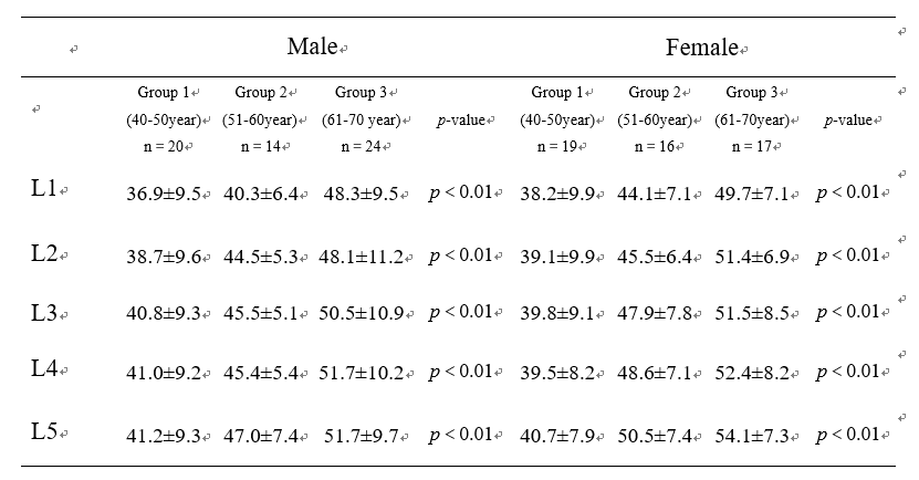
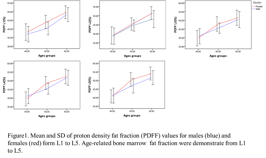
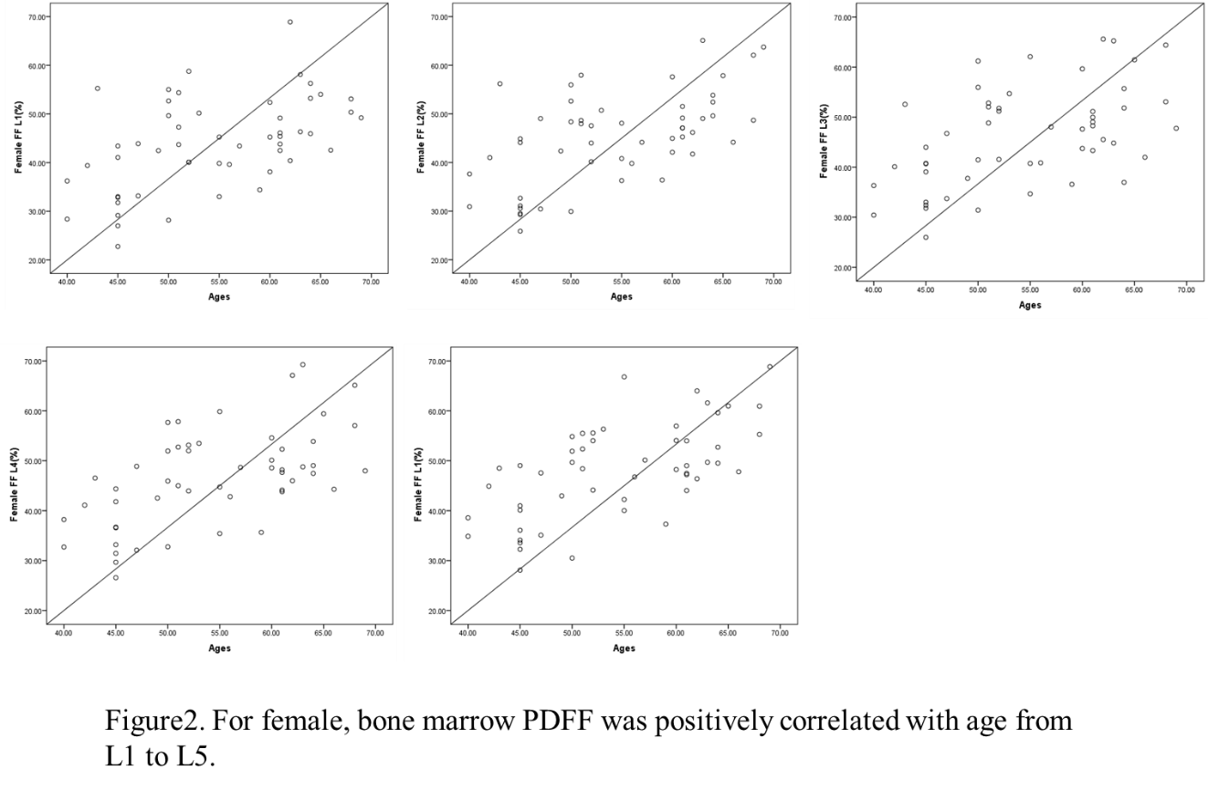
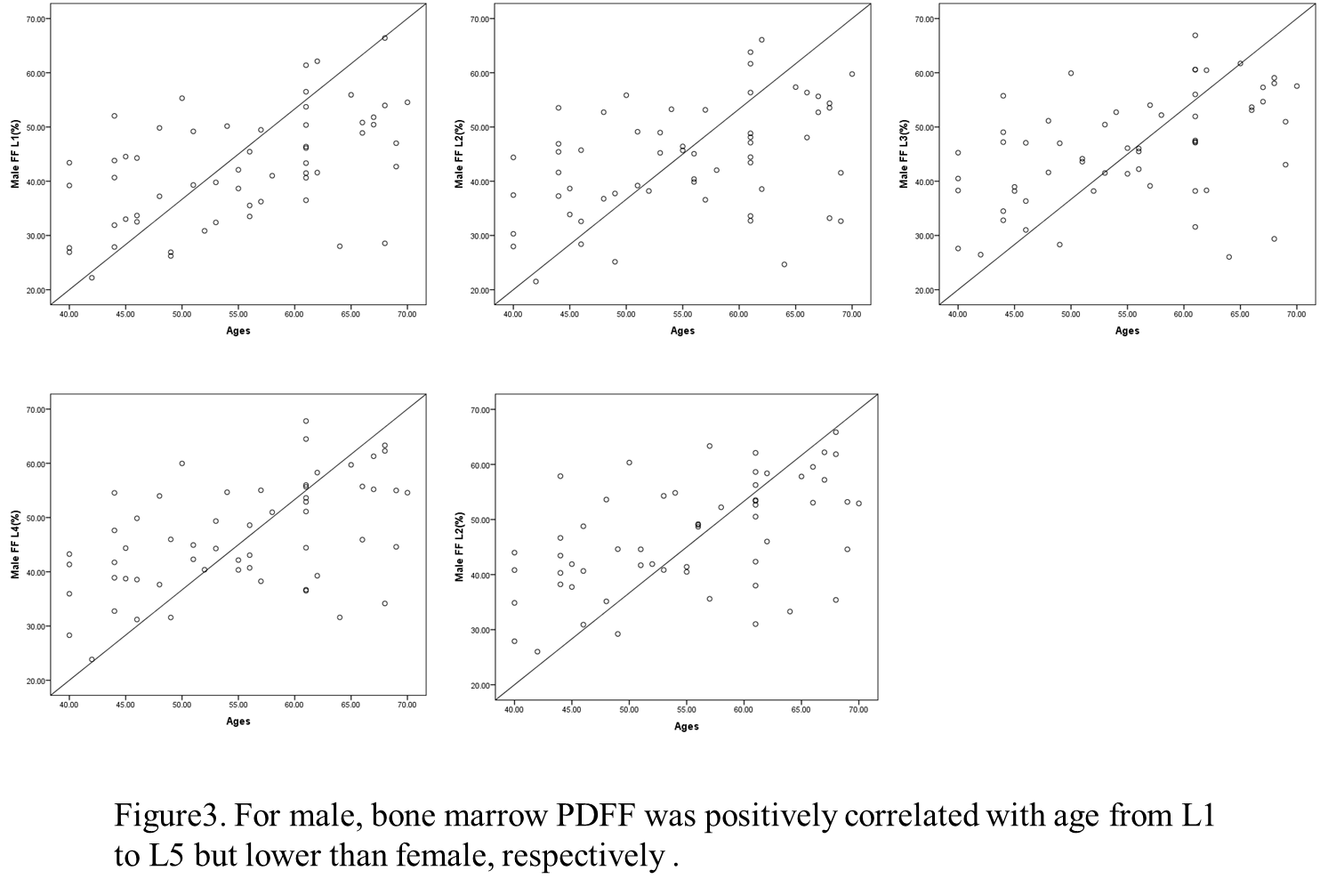
Table 1 shows the lumbar vertebral bone marrow PDFF from L1 to L5 in different age groups of male and female subjects. The values of lumbar vertebral bone marrow PDFF were significantly different (p<0.01) across three age groups from L1 to L5 in both genders. We found a higher lumbar vertebral bone marrow PDFF as the age increased in all conditions (Figure 1). Values of PDFF were generally higher in female than in male except for the PDFF of L3–L5 in the group 1 (40–50 years), while there were no significant differences of bone marrow PDFF between male and female in all groups (p>0.05). The correlations between PDFF and age in female and male subjects were shown in Figures 2 and 3, respectively. There were moderate positive correlations between bone marrow PDFF and age from L1 to L5 in both genders, while the correlations were higher in female than in male.Discussion
The most important finding of this study was that we found a age-related increase of lumbar vertebral bone marrow PDFF using MR IDEAL-IQ technique. The age-related increase of PDFF were found in both genders, which have been reported to have an anatomical changes of the lumbar vertebral bone marrow composition6-7. Limited by the development of MRI technology, in the last two decades, MRS is the main way to evaluate fat content in vertebral bone marrow using a single-voxel measurement with a long scanning time. Comparing to single-voxel MRS, IDEAL-IQ sequence provides a big FOV entirely cover L1-L5 in single scanning. The benefits of IDEAL-IQ in measuring of PDFF have been demonstrated against MRS in previous studies8-9. This study demonstrated that PDFF of vertebral bone marrow is related to age and sex. This finding corroborated with previous researches3. The differences of PDFF from L1 to L5 in both genders may help to explain the relationship between physiological changes and menopause of hormonal variation.Acknowledgements
No acknowledgement found.References
1.Rosen CJ, Ackert-Bicknell C, Rodriguez JP, et al. Marrow fat and the bone microenvironment: developmental, functional, and pathological implications. Crit Rev Eukaryot Gene Expr. 2009;19(2):109-24.
2.Paccou J, Hardouin P, Cotten A, et al. The role of bone marrow fat in skeletal health: usefulness and perspectives for clinicians. J Clin Endocrinol Metab. 2015;100(10):3613-21.
3.Li X, Kuo D, Schafer AL, et al. Quantification of vertebral bone marrow fat content using 3 Tesla MR spectroscopy: reproducibility, vertebral variation, and applications in osteoporosis. J Magn Reson Imaging. 2011;33(4):974-9.
4.Agrawal K, Agarwal Y, Chopra RK, et al. Evaluation of MR spectroscopy and diffusion-weighted MRI in postmenopausal bone strength. Cureus. 2015;7(9):e327.
5.Yeung DK, Grifth JF, Antonio GE, et al. Osteoporosis is associated with increased marrow fat content and decreased marrow fat unsaturation: a proton MR spectroscopy study. J Magn Reson Imaging. 2005;22(2):279-85.
6.Liu Y, Tang GY, Tang RB, et al. Assessment of bone marrow changes in postmenopausal women with varying bone densities: magnetic resonance spectroscopy and diffusion magnetic resonance imaging. Chin Med J (Engl). 2010;123(12):1524-7.
7.Ruschke S, Pokorney A, Baum T, et al. Measurement of vertebral bone marrow proton density fat fraction in children using quantitative water-fat MRI. MAGMA. 2017;30(5):449-60.
8.Oriol A, Valverde D, Capellades J, et al. In vivo quantifcation of response to treatment in patients with multiple myeloma by 1H magnetic resonance spectroscopy of bone marrow. MAGMA. 2007;20(2):93-101.
9.He J, Fang H, and Na Li X. Vertebral bone marrow diffusivity in normal adults with varying bone densities at 3T diffusion-weighted imaging. Acta Radiol. 2018;59(1):89-96.
Figures