1306
Value of zero echo time imaging and CT in diagnosis of bone destruction of bone tumors and tumor-like lesions1Radiology, Shanghai General Hospital, Shanghai, China, 2GE Healthcare, Shanghai, China
Synopsis
The purpose of the study was to evaluate the clinical applicability of zero echo time (ZTE) MR and compare the image quality between CT and ZTE imaging of bone tumor and tumor-like lesions. Thirty-six patients including 18 males and 18 females were recruited to undergo ZTE MR and CT. Agreement was assessed between raters and Weight Kappa statistics were performed. The difference of image quality between ZTE and CT imaging were not significant. Our results confirm that ZTE MR imaging provides accurate imaging of bone morphology with CT-like contrast that is not available with standard MR sequences.
Purpose
Bone tumor and tumor-like lesions require comprehensive imaging containing soft tissue and bone structures. Traditional MR imaging provides superior soft-tissue contrast but depicts cortical bone as signal void because of bone’s inherent short T2 relaxation time. Frequently, information for diagnosis or surgical planning requires patients to undergo multiple cross-sectional examinations (eg, X-ray plain film, CT and MR) to assess soft-tissue stabilizers and osseous support of bone tumor and tumor-like lesions. Clinically, patients with bone neoplastic lesions usually take X-ray plain film to obtain the whole bone lesions imaging. Then they usually take CT to observe the local performance of bone destructions. Furthermore, they undergo MR to show the suffered soft tissue around the bone tumor lesions. Therefore, there are too many examinations for the patients with bone neoplastic lesions. Zero echo time (ZTE) can acquire FID signals directly after the RF pulse.1 Inverse-logarithmic rescaling of ZTE images provides contrast between soft tissue and bone comparable to that of CT.2-3 We sought to apply ZTE to skeletal imaging of bone tumor and tumor-like lesions.Methods
This prospective study was approved by the Institutional Ethical Committee of our hospital. We recruited the patients with bone tumor and tumor-like lesions between December 2017 and May 2018 in our hospital. Among 36 patients, 18 were males (35±20 years old) and 18 were females (42±20 years old). All patients underwent biopsy to acquire pathology results. CT was performed by using a GE Discovery CT750 HD scanner (n = 30) or a GE LightSpeed VCT scanner (n = 6) (GE Healthcare, Milwaukee, USA). The scan parameters were as follows: 0.625mm Section thickness, 14–29cm field of view, 0.273–0.559mm pixel size and 512*512 matrix size. MR imaging included coronal inversion-recovery and coronal, sagittal, and axial fast spin-echo proton density–weighted acquisitions. ZTE images were acquired using a 3.0-T MR750 scanner (GE Healthcare, Milwaukee, USA) with the following parameters: 1mm section thickness, 32cm field of view and 320x320 Matrix size. The total acquisition time was 4 minutes. Two board-certified musculoskeletal radiologists with more than five years of experience scored CT and ZTE images. CT and ZTE images scans were coregistered together and randomly reviewed using a 5-point grading scale: 1. Very poor: Obscured anatomic detail, not sufficient for diagnosis; 2. Poor: No clear anatomic details or anatomic details are not sufficiently detectable; 3. Fair: Most anatomic structures are sufficient for diagnosis, but some pictures are inadequate for evaluation; 4. Good: Anatomic structures and details are present in a level that allows proper but not excellent evaluation of the images; 5. Excellent: Distinct anatomic detail leading to clear and easy evaluation. The statistical analysis was carried out using Mann–Whitney test and Weight Kappa statistics . Interpretation of agreement between raters was based on published standards: 0.00–0.20 indicated slight; 0.21–0.40, fair; 0.41–0.60, moderate; 0.61–0.80, substantial; and 0.81–1, perfect agreement.Results
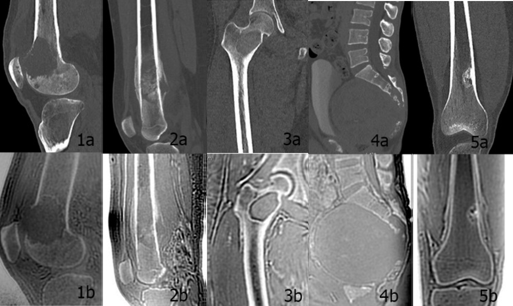
There were thirteen types of bone tumors and tumor-like lesions in 36 patients, including eight cases of osteosarcoma, eight cases of metastases, four cases of chondrosarcoma, three cases of giant cell tumor, three cases of fibrous dysplasia, two cases of schwannoma, two cases of Ewing's sarcoma, one case bone cyst, one case of synovial sarcoma, one case of liposarcoma, one case of non-ossifying fibroma, one case of tubular sarcoma and one case of chordoma. Both of ZTE and CT images show the changes of bone destruction and periosteal reaction of bone tumors, which can be used for clinical diagnosis (Fig.1). There was no significant difference between CT and ZTE MR (P = 0.067). The consistency analysis between the two reviewers in CT group and MR group showed good consistency (K = 1, K = 0.84).Discussion and Conclusion
In summary, ZTE-MR imaging is another breakthrough in the field of MR compared with conventional T1, T2 and proton density weighted imaging. This study provides a new scanning scheme ZTE, which is similar to the contrast of CT images. The results show that ZTE-MR meets the diagnostic requirements. As an alternative to CT, ZTE-MR can help patients with bone tumors to simplify the inspection process.Acknowledgements
Contract grant sponsor: Biomedical Engineering Cross-Research Fund of Shanghai Jiaotong University; Contract grant number: YG2017QN25.References
1. Weiger M, Hennel F, Pruessmann KP. Sweep MRI with algebraic reconstruction. Magn Reson Med 2010;64:1685–1695.
2. Breighner RE, Endo Y, Konin GP, et al. Technical developments: zero echo time imaging of the shoulder: enhanced osseous detail by using MR imaging. Radiology 2018;286:960–966.
3. Dreher W, Bardenhagen I, Huang L, et al. On the suppression of background signals originating from NMR hardware components. Application to zero echo time imaging and relaxation time analysis. Magn Reson Imaging 2016;34:264–270.
Figures