1305
Research on the feasibility of MR imaging of patients receiving anterior cervical surgery using MAVRIC SL-STIR sequence at 3T1Renmin Hospital of Wuhan University, Wuhan, China, 2GE Healthcare, Beijing, China
Synopsis
Metal implants are now very common in modern joint and spine surgeries. However, conventional MR images are significantly compromised by implant-induced magnetic susceptibility artifacts. A novel metal artifacts reduction technique, termed MAVRIC SL was proposed. The purpose of this study is to evaluate its clinical feasibility and diagnostic value in patients after anterior cervical surgery compared with routine 2D FSE sequence at 3T. As a result, although the image quality of MAVRIC SL is limited at 3T, it can still provide important additional diagnostic information through substantially reduced metal artifacts.
Introduction
Metal implants are now very common in modern joint and spine surgeries. However, conventional MR images are significantly compromised by implant-induced magnetic susceptibility artifacts, which will limit the accuracy of image interpretation.1, 2Minimizing metal artifacts can offer great helps to better depict the metallic implants and surrounding anatomic structures. Meanwhile, uniform fat suppression is beneficial to improve detection rate of lesions, such as tumor, bleeding, edema, infection and so on3. Therefore, minimizing metal artifacts and acquiring optimal effect of fat suppression are crucial for post-surgery evaluation of patients with fixed metallic implants. Lately, a novel MRI technique, termed MAVRIC SL (Multi-Acquisition with Variable Resonance Image Combination SeLective) was proposed, which combines the slice-encoding metal artifact correction (SEMAC) with Multi-Acquisition with Variable Resonance Image Combination (MAVRIC). This technique shows a significant advantage on metal artifacts reduction4. Furthermore, MAVRIC SL-STIR combines the 180o inversion pulse with MAVRIC SL for fat suppression. The purpose of this study is to evaluate the clinical feasibility and diagnostic value of MAVRIC SL-STIR in patients after anterior cervical surgery compared with routine 2D FSE sequence at 3T MRI.Methods
Institutional review board approval and informed consent were obtained for this study. Images were acquired on a 3T MR scanner (GE Medical Systems, Milwaukee, WI). In all, 15 patients (9 males and 6 females, average age: 53.6±14.5) after anterior surgery from 5 days to 6 years were included in this study. Besides the routine cervical spine protocols, an additional MAVRIC SL-STIR sequence was scanned in sagittal plane with similar spatial resolution to compare with the routine 2D FSE sequence (bandwidth optimized STIR). For quantitative evaluation, the areas of metal artifacts regions were manually outlined and measured in the mid-sagittal plane by the same radiologist for the both sequences (MARVRIC SL-STIR and bandwidth optimized STIR). Statistically comparison of the areas of metal artifacts between the two sequences were conducted using a One-way ANOVA test. For qualitative evaluation, two musculoskeletal radiologists separately analyzed the image quality including geometric image distortion, blurring, noise and uniformity of fat suppression using a five-point scale (1, severe artifacts and non-diagnostic; 5, nearly no artifacts and definitively diagnostic).Besides, the visibility of three anatomic structures including the pedicle, vertebral body and dural sac near the implants were also assessed using a five-point scale (1, anatomic structure not visible; 5, good delineation of anatomic structure). Comparison between the two sequences was performed using a Kruskal-Wallis H test .Results
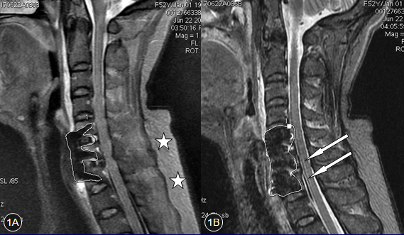
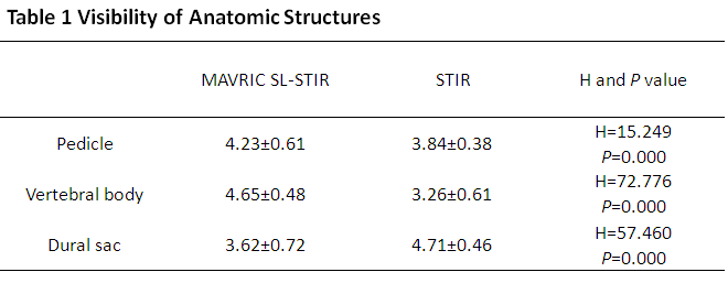
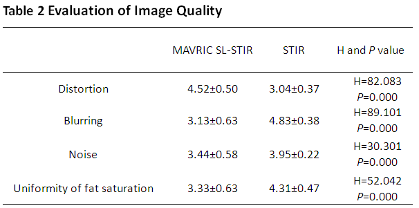
Respective advantages were acquired by the two sequences (Fig.1).The mean areas of metal artifacts regions for MAVRIC SL-STIR and bandwidth optimized STIR images were 428.467mm2 and 800.200mm2, respectively. The area of metal artifacts was significantly reduced on MAVRIC SL-STIR images (P=0.002). Table 1 shows the comparison results of image quality scores. MAVRIC SL-STIR demonstrated superior image distortion (P<0.001), while blurring was less in STIR images. Table 2 summarizes the visibility scores of anatomic structures for comparison between the two sequences. MAVRIC SL-STIR acquired better visibility of the pedicle and vertebral body (P <0.001). However, the visibility of the dural sac for MAVRIC SL-STIR images were decreased (P <0.001).Discussion and Conclusion
As shown in the results, MAVRIC SL-STIR can effectively reduce the metal artifacts and improve delineation of the metallic implants compared to the routine 2D FSE sequence (bandwidth optimized STIR). But it suffers from image blurring which mainly caused by long echo train length (ETL=20) and excitation mode of RF pulse.4In addition, the relative long acquisition time (about 11 minutes) may bring a risk of motion artifacts. Moreover, the effect of fat suppression of MAVRIC SL-STIR was reduced compared to STIR technique. In conclusion, although the image quality of MAVRIC SL-STIR sequence is limited at 3T, its clinical application for patients after anterior cervical surgery is feasible.Acknowledgements
No acknowledgement found.References
1.Viano, A. M., Gronemeyer, S. A., Haliloglu, M. and Hoffer, F. A., Improved MR imaging for patients with metallic implants, Magnetic Resonance Imaging, 2000, 18(3):287-295.
2.Hargreaves, B., Worters, P. W., Pauly, K. B., Pauly, J. M., Koch, K. M. and Gold, G. E., Metal Induced Artifacts in MRI, Ajr American Journal of Roentgenology, 2011, 197(3):547-555.
3.Rutherford, E. E., Tarplett, L. J., Davies, E. M., Harley, J. M. and King, L. J., Lumbar spine fusion and stabilization: hardware, techniques, and imaging appearances, Radiographics A Review Publication of the Radiological Society of North America Inc, 2007, 27(6):1737-1746.
4.Koch, K. M., Brau, A. C., Chen, W., Gold, G. E., Hargreaves, B. A., Koff, M. and Mckinnon, G. C., et al., Imaging near metal with a MAVRIC‐SEMAC hybrid, Magnetic Resonance in Medicine Official Journal of the Society of Magnetic Resonance in Medicine, 2011, 65(1):71-82.
Figures