1297
Estimating the diagnostic value of IDEAL-IQ for vertebral compression fractures caused by osteoporosis and metastasis1Department of Radiology, WeiFang Traditional Chinese Hospital, Wei Fang, Shandong,P.R. China, China, 2WeiFang Traditional Chinese Hospital, Wei Fang, Shandong,P.R. China, China, 3GE Healthcare, MR Research China, Beijing, P.R. China, China
Synopsis
Traditional imaging methods are challenging to diagnose the vertebral compression fractures caused by osteoporosis and metastasis. In this study, the iterative decomposition of water and fat with echo asymmetry and the least squares estimation quantification sequence (IDEAL-IQ)technique, as a novel fat quantification technique, was applied to quantitatively determine the bone marrow fat content for the patients with osteoporosis caused and metastasis caused vertebral compression fractures. We found that the fat fraction (FF) and FF ratio of bone marrow fat in the vertebral body lesions of metastasis were significantly reduced compared with the acute compression fractures due to osteoporosis. Therefore, IDEAL-IQ has been proven an effective method for quantitative diagnosis of vertebral compression fractures.
Abstract
Introduction
Vertebral compression fractures in elder population are mainly caused by osteoporosis and metastasis. Clinical diagnosis are usually accomplished based on the morphological changes occurred in the vertebral compression fractures, through X-ray, computed tomography or conventional magnetic resonance imaging (MRI). While they performed effectively in some cases, these imaging techniques still remain challenging in the identification of most lesions1. The iterative decomposition of water and fat with echo asymmetry and the least squares estimation quantification sequence (IDEAL-IQ) is a novel fat quantification technique, but has rarely been reported to assess the bone marrow fat content in vertebral lesions.
Therefore, this study aimed to investigate the feasibility of IDEAL-IQ in the quantitative estimation of the bone marrow fat content in osteoporosis and metastasis caused vertebral compression fractures, and to explore if the quantified fat relevant results can serve as biomarkers to distinguish the bone fractures with different causes.
Materials and Methods
Subjects
Seventy patients (mean age:67.15±16 years old) with acute vertebral compression fractures caused by osteoporosis(n=35) or metastasis(n=35) were recruited in this study for MRI experiments. Written informed consent was obtained from each patient.
MRI experiments
All experiments were performed at GE 3.0 T Silent 750W with posterior array coils, Routine MRI and IDEAL-IQ technique were applied. For IDEAL-IQ measurement, the scan parameters included: repetition time=5.5ms, echo time=2.6ms NEX=3, field-of-view=32x32cm, slice thickness=5mm, phase number=160, gap between slices=0.5mm and flip angle=3°. The total scan time was 1 minutes 26 seconds. The parametric fat fraction and R2* mapping were then automatically obtained.
Data analysis
All data were analyzed at a GE MR workstation (Advantage workstation 4.6; GE Medical Systems). On the acquired T1- and T2- weighted images, two experienced radiologists were employed to independently measure the signal intensities in the lesions of vertebral compression fractures for all patients. The corresponding FF and R2* values of lesion regions were also obtained from FF and R2* maps. In addition, a FF ratio, defined as FF value of the lesion / FF value in the normal vertebral bone marrow, was calculated for each patient.
In SPSS software version 17.0, the embedded Mann-Whitney U test was used to determine the difference between the parameters of osteoporosis and metastasis, including FF, FF ratio and R2* . In addition, the receiver operating characteristic curve (ROC) analysis was applied to estimate the diagnostic efficacy of FF, FF ratio and R2* in vertebral compression fractures. P<0.05 was considered statistically significant.
Results
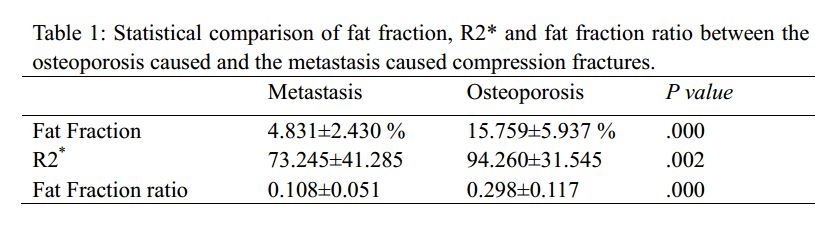
In Table 1, significant lower FF values, R2* values and FF ratios of bone marrow fat content were all found (P<0.05) in the metastasis caused vertebral lesions than the osteoporosis caused acute compression fractures (FF:4.831±2.430% vs 15.759±5.937 %; R2*:73.245±41.285 vs 94.260±31.545; FF ratio: 0.108±0.051 vs 0.298±0.117).
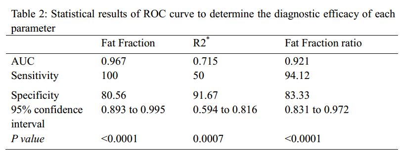
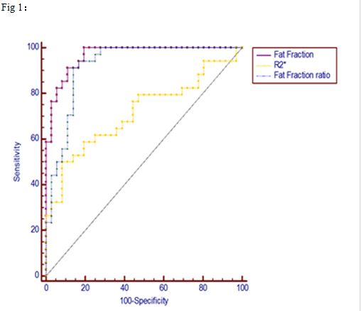
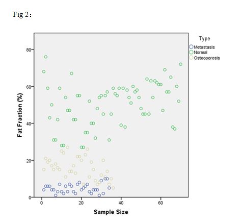
High AUC values of FF, R2*, and FF ratio were shown, indicating a robust differentiation of metastatic tumor compression fractures from osteoporosis acute compression fractures ( 0.967, 0.715, and 0.921; Table2). The ROC curve analyzed the specificity and sensitivity of different diagnosis (figure 1).The scatter diagram directly reflects the distribution of fat fraction in the vertebral lesions caused by normal vertebral metastases and osteoporosis (figure 2).
Discussion
In this study, IDEAL-IQ technique was used to quantitatively assess the vertebral compression fractures with different causes. The resultant FF and FF ratio showed significant lower values in the vertebral body with metastasis than the acute compression fracture due to osteoporosis. This is because the metastasis as tumor cell reaches the vertebrae by blood and grows invasively, replacing normal bone marrow tissues. Meanwhile, the tumor cells show focal growth leading to the destruction of the bone tissue at the metastasis and the bone marrow edema surrounded. Both effects lead to a significantly reduced adipose tissue in the vertebral body. In comparison, osteoporosis caused vertebral compression fracture occurs under the action of slight external force. The internal vertebral body in the early stage of fracture is mainly due to the bone marrow edema caused by compression fracture and small amount of bleeding. Compared with the vertebral lesion area with metastatic tumor, the bone marrow fat and water content increases in the osteoporosis acute compression fracture 2-3. Therefore, through quantitative assessment of fat fraction in vertebral lesions, IDEAL-IQ reflected the dynamic alterations of fat and water content in bone marrow caused by the above mentioned pathological factors.
Conclusion
In conclusion, IDEAL-IQ has revealed its feasibility and clinical values in distinguishing the osteoporosis caused from the metastasis caused vertebral compression fractures, and can thus be routinely applied in the clinic.
Acknowledgements
Thanks to colleagues who worked together in the study.References
1. Dong Hyun Kim,Hye Jin Yoo, et al. Differentiation of Acute Osteoporotic and Malignant Vertebral Fractures by Quantification of Fat Fraction With a Dixon MRI Sequence. AJR, 2017, 209(9):1331–1339.
2. Dimitrios C. Karampinos, Stefan Ruschke, et al. Quantitative MRI and Spectroscopy of Bone Marrow. J. Mang. Reson. Imaging 2018;47:332–353.
3. Frederic Carsten Schmeel,Julian Alexander Luetkens, et al. Proton density fat fraction (PDFF) MRI for differentiation of benign and malignant vertebral lesions. Eur Radiol ,2018,28:2397–2405.
Figures