1287
Perfect In-Phase Zero TE for Musculoskeletal Imaging1GE Healthcare, Stockholm, Sweden, 2GE Healthcare, Munich, Germany, 3GE Healthcare, San Diego, CA, United States, 4GE Healthcare, Waukusha, WI, United States
Synopsis
Large FOV Zero Echo-Time (ZTE) has been challenging due to chemical shift artifacts, caused primarily by fat water dephasing, for low readout band-widths (rBW). To correct for this Perfect In-Phase ZTE (pipZTE) is proposed where the chemical shift artifact is removed by acquiring data from multiple rBWs, and then separating the signal into an in-phase and off-resonance compartment in the reconstruction. In this work we explore the performance and properties of the pipZTE approach when scanning large FOV and demanding subjects.
Introduction
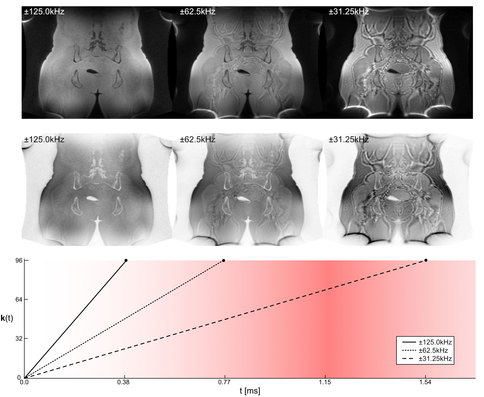
In radiotherapy (RT) planning and PET/MR attenuation correction proton density (PD) weighted Zero Echo-Time (ZTE) acquisition strategies have received a prominent role over the last years (1). This is primarily due to its ability of detection MR bone signals and differentiating them from surrounding soft-tissue and air. For common post-processing procedures such as bone enhancement (2) and pseudo-CT generation (1) a high soft-tissue uniformity is crucial. Despite a nominal echo time of TE = 0, ZTE images can still contain significant chemical shift induced out-phase effects and thereby significantly degrade soft-tissue appearance, especially at fat-water tissue interfaces. This is because the ZTE readout is of finite duration and distant k-space points are sampled at effective echo times which can approach fat-water out-phase echo time (TEout-phase~1.15ms at 3T) (cf. Fig. 1).
$$data(\mathbf{k}_{n})= \\ \int_{}^{}d^{3}r\begin{bmatrix}image_{in-phase}(\mathbf{r})\frac{e^{i\omega_{water}} {TE}_{n}+e^{i\omega_{fat}} {TE}_{n}}{2}+image_{out-phase}(\mathbf{r})\frac{e^{i\omega_{water}} {TE}_{n}-e^{i\omega_{fat}} {TE}_{n}}{2}\end{bmatrix}e^{i\mathbf{k}_{n}\mathbf{r}}\\= image_{in-phase}(\mathbf{k}_{n})\frac{e^{i\omega_{water}} {TE}_{n}+e^{i\omega_{fat}} {TE}_{n}}{2}+image_{out-phase}({\mathbf{k}_{n}})\frac{e^{i\omega_{water}} {TE}_{n}-e^{i\omega_{fat}} {TE}_{n}}{2}$$
In this work we present a novel method, called perfect in-phase ZTE (pipZTE), which produces ZTE images clean of out-phase disturbances via a k-space based in-phase/out-phase chemical shift decomposition. The pipZTE method is investigated in terms of spatial resolution and SNR characteristics and demonstrated for musculoskeletal imaging and whole-body bone imaging.
Methods
ZTE imaging was performed based on a modified Rotating Ultra-Fast Imaging Sequence (RUFIS) (5). In order to allow magnetization preparation (such as IR preparation, and T2 preparation) the ZTE readout is divided into segments, with each segment containing a certain number of spokes per segment. In pipZTE each segmented gets repeated with the readout gradient being scaled providing k-space samples (i.e. data(kn)) acquired at different effective echo times (TEn=n*Δt, where Δt ithe sampling time).
Assuming a fat-water shift of 3.4ppm each k-space sample can then be reconstructed into two compartments (in-phase and off-resonance) (Eq. 1), using either regularized pseudo-inversion or truncated single value decomposition (SVD). With the first k-space samples acquired at TE ~0 the off-resonance effects become increasingly destructive approaching ~1.15ms at 3.0T. Data reconstruction was performed on the scanner using ORCHESTRA (ORCHESTRA, GE Healthcare, Chicago, IL) and DICOM images were automatically inserted into the scanner database. Healthy volunteers were scanned on a 3.0T clinical MRI system (SIGNA Premier, GE Healthcare, Chicago, IL) using a 60ch AIR technology receive coil (AIR AA+PA, GE Healthcare, Chicago, IL) with relevant scan parameters being FOV 320mm, resolution 1.5x1.5x1.5mm3, rBW ±62.5/50.0kHz, NEX 1, flip angle 0.8°, and a total scan time of 3:42min. Scan time and resolution matched ZTE data was acquired in addition for comparison.
Results and Discussoion
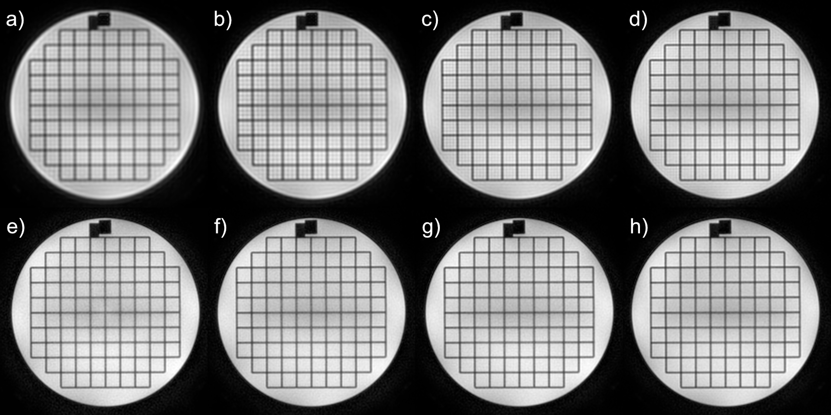
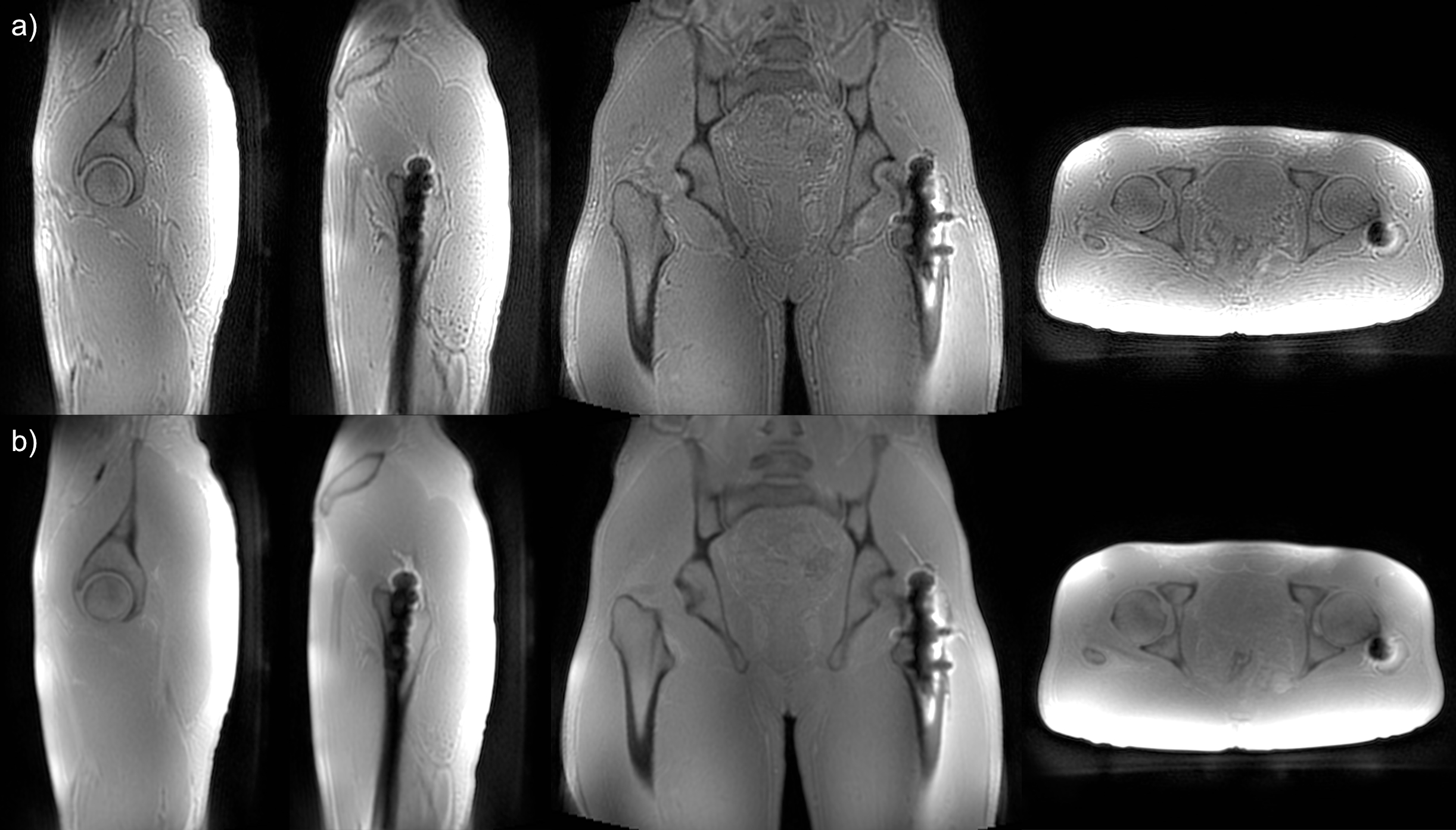
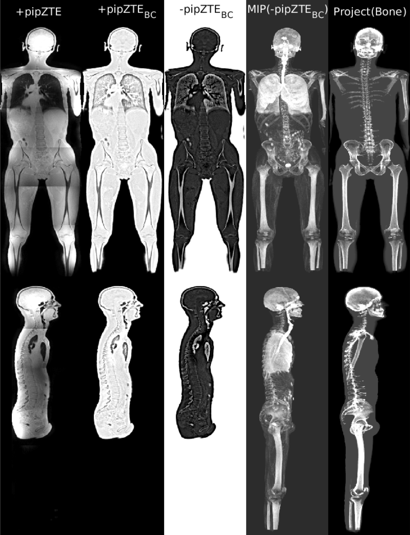
In Figure 2 we show the performance of regularized pseudo-inverse in comparison to SVD as a method for signal decomposition. While the regularized pseudo-inverse is capable of resolving more information at high gradient scaling factors, the SVD performs remarkably good retaining the resolution, even for gradient scaling levels of 50%. In Figure3 a SNR improvement, in favor of the pipZTE, can be seen directly in the images. We attribute this improvement to the constructive signal composition of the In-Phase compartment where destructive off-resonance species are effectively removed. On the top left panel (Figure 3a) clear chemical shift artifacts can be seen in the ZTE data as a result of water-fat dephasing at specific spatial frequencies. In the top right panel (Figure 2b) pipZTE data is presented showing no apparent chemical shift artifacts, but an improved soft-tissue signal homogeneity. A slight resolution loss can be seen for the pipZTE data, in comparison with ZTE, as a result of the reconstruction. Optimal scan parameters will have to be investigated per anatomy, along with gradient scaling scheme. A two-point approach has been found adequate for the data presented in this work, but for challenging areas or inclusion of multiple off-resonance peaks an extended gradient scaling scheme might be beneficial. Figure 4 shows whole body pipZTE images in coronal (top) and axial (bottom) view before (1st column) and after bias correction (2nd column). Inverting the gray scale emphasize the bone structures in a way similar to CT (3rd column). A maximum intensity projection (MIP) of low ZTE signals (originating from either air or bone) within the body mask is illustrated in the 4th column. Air cavities (i.e. the lungs, trachea, abdominal gas) can effectively be removed via connected component analysis (5th column).Conclusion
In summary, we have presented a novel perfect in-phase ZTE (pipZTE) method, which effectively removes fat-water out-phase signal disturbances via k-space based in-phase / out-phase chemical shift decomposition. The obtained images demonstrate exceptional soft-tissue uniformity as required for musculoskeletal imaging and in particular bone imaging and pseudo CT conversion.Acknowledgements
No acknowledgement found.References
- Wiesinger F, et.al., Zero TE-based pseudo-CT conversion in the head and its application in PET/MR attenuation correction and MR-guided radiation therapy planning. Magn Reson Med. 2018 0ct;80(4):1440-51
- Breighner RE, et.al., Zero Echo Time imaging of the Shoulder: Enhanced Osseous Detail by Using MR Imaging. Radiology. 2018 Mar;286(3):960-6
- Wiesinger F, et.al., Whole Body Skeletal Imaging Using Zero TE. In: Proceedings of the 24th Annual meeting of ISMRM. Singapore, Singapore, 2016. p. 675
- Engström M, Cozzini C, Wiesinger F, Perfect In-Phase ZTE for improved MR Attenuation Correction. In: Proceedings of the 7th Conference on PET/MR and SPECT/MR. Elba, Italy, 2018.
- Madio DP, Lowe IJ. Ultra-fast imaging using low flip angles and fids. Magn Reson Med. 1995;34:525-529
Figures