1285
Ultrashort echo time MRI (UTE-MRI) quantifications of cortical bone varied between scans at room temperature and body temperature1Radiology, University of california, San Diego, San Diego, CA, United States, 2Radiology Service, VA San Diego Healthcare System, San Diego, CA, United States
Synopsis
Several quantitative ultrashort echo time MRI (UTE-MRI) techniques have recently been employed to assess cortical bone microstructure. Such techniques were examined mostly ex vivo at room temperature and demonstrated strong correlations with bone microstructure as measured with micro computed tomography (μCT). However, MRI properties of cortical bone may differ in vivo due to higher temperature. We have investigated several UTE-MRI quantifications of cortical bone at body and room temperatures. Significant variations of bone UTE-MRI measures were observed between room and body temperatures. Implementing a linear correction method on UTE-MRI measures based on the presented results here might improve the validity of the techniques for in vivo studies.
Introduction
Quantitative ultrashort echo time MRI (UTE-MRI) is attracting more attention from orthopeadic research groups for cortical bone assessment. Several UTE techniques have been examined ex vivo and demonstrated strong correlations with bone microstructure as measured with micro computed tomography (μCT)(1–4). Ex vivo experiments routinely were performed at room temperature yet the target bone for future clinical studies resides in body temperature. As reported in the literature, MRI properties in cortical bone are functions of the temperature (5–7). Consequently, concluded performance level of UTE-MRI techniques based on ex vivo results cannot be expected for in vivo studies without considering the temperature effects. However, the differences between in vivo cortical bone and ex vivo specimens are not limited to the temperature. This study aimed to investigate variation of UTE-MRI quantifications of cortical bone between scans at room temperature and scans at body temperature. Studied UTE techniques included single- and bi-component T2* fittings, inversion recovery UTE (IR-UTE) T2*, T1, and two-pool MT modeling.Methods
Sample preparation: Cortical bone specimens were harvested from fourteen human tibial and femoral midshafts (63±21 years old, 6 females). Specimens were cut in 30 mm length using a commercial band saw. A rectangular strip was excised from each specimen using a low-speed diamond saw (Isomet 1000, Buehler, IL). The final approximate dimensions of bone strips were 4×2×30 mm.
Forced air regulated temperature device: An MR compatible device was designed and manufactured in-house to force a directed air flow with a regulated temperature on the specimens during the MRI scans.
UTE-MRI sequences: Bone specimens were immersed in phosphate buffered saline (PBS) for twelve hours before scans. All specimens were placed in a 30-mL syringe filled with perfluoropolyether (Fomblin, Ausimont, Thorofare, NJ) to minimize dehydration and susceptibility artifacts. The UTE-MRI scans were performed on a 3T clinical scanner (MR750, GE Healthcare Technologies, WI) using home-made 1-inch diameter transmit/receive birdcage coil. Scans were performed first at body temperature (i.e., 37.5±2˚c) and second at room temperature (19±1˚c). The quantitative UTE-MRI protocol involved A) five sets of dual-echo 3D-UTE-Cones sequences (TR=24.3, TEs=0.032, 0.2, 0.4, 0.8, 2.2, 4.4, 6.6, 8.8, 11 and 15ms) for T2* single- and bi-component analyses, B) five set of 3D-IR-UTE-Cones sequence (TI=45, TR=100, TEs=0.032, 0.2, 0.4, 0.6 and 1ms, FA=20˚) for IR-T2* measurements, C) an actual FA variable TR (AFI-VTR) sequence (AFI: TE=0.032, TRs=20ms and 100ms, VTR: TE=0.032, TRs=20-100ms, FA=45˚) for T1 measurements (8), and D) a set of 3D-UTE-Cones-MT sequences (MT saturation pulse power=400°, 600°, and 800°, frequency offset=2, 5, 10, 20, and 50kHz, FA=10˚) for two-pool MT modelling (9). Field of view (FOV), matrix dimension and slice thickness were 40mm, 160×160 and 3mm, respectively.
Data analysis: Average UTE-MRI quantifications (T2*, T1, and MT modeling) were calculated for each scanned bone specimen at room and body temperatures. Statistical differences were calculated between results at room and body temperatures using the two-tailed paired Student’s t-test. P-values below 0.05 were considered significant. All measurements and models were performed using in-housed developed codes in MATLAB (The Mathworks Inc., Natick, MA, USA).
Results
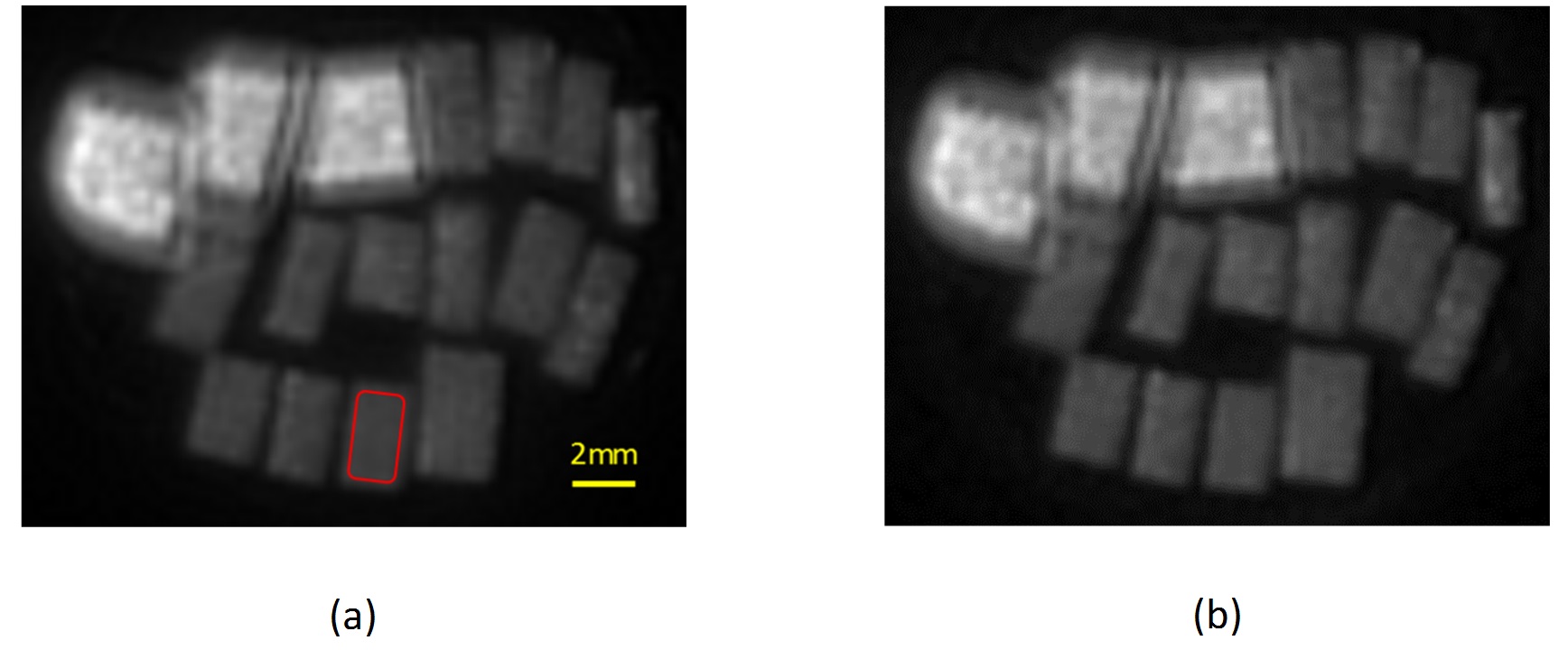
Figure 1 shows the UTE-MRI images of the
fourteen studied cortical bone specimens at room and body temperatures. No
clear differences were observed between images. Figures
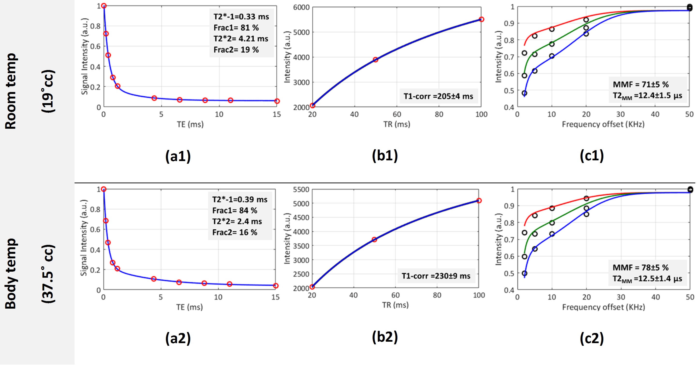
2a-c show the bi-component T2* fitting, single-component T1 recovery fitting
and the two-pool MT modeling analyses, respectively, for a representative bone
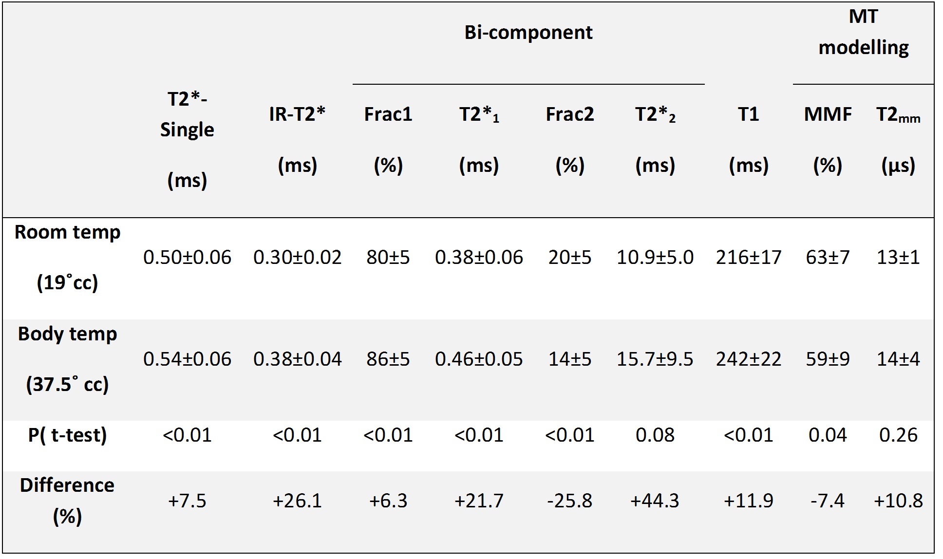
specimen (54-year old male) at two studied temperatures. Figure 3 (Table)
presents average values of measured UTE-MRI biomarkers at room and body
temperatures in addition to their statistical differences. All UTE-MRI measures except T2mm demonstrated
significant differences between room and body temperatures (P<0.05).Discussion
Significant variations of UTE-MRI measures between room and body temperatures suggest that the conclusions of ex vivo studies need to be modified before extending to in vivo studies. T2*1, T2*2 and IR-T2* were significantly higher at body temperature compared with room temperature (>20%, P<0.01). Variation of bi-component fractions indicates different extents of temperature impacts on the bound and pore water pools. Higher T1 in body temperature agreed with the reported results in the literature (6).Conclusion
This study highlighted the impact of temperature differences between ex vivo and in vivo studies on cortical bone, even though such differences are not limited to the temperature. Significant temperature-based differences in UTE-MRI measures suggest that the conclusions of ex vivo studies need to be modified before applying them to in vivo studies. Implementing a linear correction method on UTE-MRI measures based on the presented results here might improve the validity of the techniques for in vivo studies.Acknowledgements
The authors acknowledge grant support from NIH (1R21AR073496, R01AR068987) and VA Clinical Science and Rehabilitation R&D Awards (I01CX001388 and I01RX002604).References
1. Du J, Bydder GM. Qualitative and quantitative ultrashort-TE MRI of cortical bone. NMR Biomed. 2013;26:489–506. doi: 10.1002/nbm.2906.
2. Bae WC, Chen PC, Chung CB, Masuda K, D’Lima D, Du J. Quantitative ultrashort echo time (UTE) MRI of human cortical bone: Correlation with porosity and biomechanical properties. J. Bone Miner. Res. 2012;27:848–857. doi: 10.1002/jbmr.1535.
3. Rajapakse CS, Bashoor-Zadeh M, Li C, Sun W, Wright AC, Wehrli FW. Volumetric Cortical Bone Porosity Assessment with MR Imaging: Validation and Clinical Feasibility. Radiology [Internet] 2015;276:526–35. doi: 10.1148/radiol.15141850.
4. Manhard MK, Uppuganti S, Granke M, Gochberg DF, Nyman JS, Does MD. MRI-derived bound and pore water concentrations as predictors of fracture resistance. Bone [Internet] 2016;87:1–10. doi: 10.1016/j.bone.2016.03.007.
5. Ozhinsky E, Han M, Bucknor M, Krug R, Rieke V. T2-based temperature monitoring in bone marrow for MR-guided focused ultrasound. J. Ther. Ultrasound 2016;4:1–9. doi: 10.1186/s40349-016-0073-8.
6. Han M, Scott SJ, Ozhinsky E, Salgaonakar VA, Jones PD, Larson PEZ, Diederich CJ, Rieke V, Krug R. Assessing temperature changes in cortical bone using variable flip-angle ultrashort echo-time MRI. AIP Conf. Proc. 2017;1821. doi: 10.1063/1.4977625.
7. Ramsay E, Mougenot C, Kazem M, Laetsch TW, Chopra R. Temperature-dependent MR signals in cortical bone: Potential for monitoring temperature changes during high-intensity focused ultrasound treatment in bone. Magn. Reson. Med. 2015;74:1095–1102. doi: 10.1002/mrm.25492.
8. Ma Y-J, Lu X, Carl M, Zhu Y, Szeverenyi NM, Bydder GM, Chang EY, Du J. Accurate T 1 mapping of short T 2 tissues using a three-dimensional ultrashort echo time cones actual flip angle imaging-variable repetition time (3D UTE-Cones AFI-VTR) method. Magn. Reson. Med. [Internet] 2018;00:1–11. doi: 10.1002/mrm.27066.
9. Ma Y-J, Chang EY, Carl M, Du J. Quantitative magnetization transfer ultrashort echo time imaging using a time-efficient 3D multispoke Cones sequence. Magn. Reson. Med. [Internet] 2017;00:1–9. doi: 10.1002/mrm.26716.
Figures