1254
Compressed-sensing 1mm-isotropic 3D Whole-Heart Water/Fat Coronary MR Angiography in ~10 minutes1School of Biomedical Engineering and Imaging Sciences, King's College London, London, United Kingdom, 2Cardiovascular MR Predevelopment, Siemens Healthcare GmbH, Erlangen, Germany, 3MR Research Collaborations, Siemens Healthcare GmbH, Frimley, United Kingdom
Synopsis
Prolonged acquisition time and robust fat suppression remain a challenge for coronary MR angiography. Here we present a highly efficient undersampled whole-heart water/fat CMRA acquisition and reconstruction approach, for improved fat suppression at 3Twithin an overall short scan time. The proposed approach is integrated with 2D translational motion correction, to enable 100% respiratory scan efficiency, which in combination with an undersampled acquisition trajectory allows the acquisition of high-resolution water/fat datasets in ~10 minutes. The feasibility of the proposed approach was tested in healthy subjects, and results show improved depiction of the coronary arteries compared to conventional fat suppression.
Introduction
Water/fat 3D coronary MR angiography (CMRA) has shown promising results for improving contrast and reducing artefacts arising from subcutaneous and cardiac fat compared to conventional fat-suppressed 3D CMRA, particularly at higher magnetic fields1. However, acquiring multiple echoes for water/fat separation significantly increases TR, reducing the number of segments that can be acquired within the mid-diastolic rest period of the cardiac cycle, and therefore increasing scan time. Furthermore, as whole-heart volumetric coverage and high-spatial resolution are required for visualisation of the coronary anatomy, water/fat 3D CMRA acquisitions require robust mechanisms for respiratory motion compensation. Conventionally, respiratory motion-compensation schemes rely on the use of 1D diaphragmatic gating and tracking, resulting in even longer acquisition times. This prolonged and unpredictable scan duration has so far hindered the wide adoption of 3D water/fat CMRA scans in clinical routine.
Here we propose an undersampled motion-corrected water/fat CMRA approach, based on a dual-echo 3D sequence, which enables the acquisition of 1mm-isotropic whole-heart water/fat images in ~10 minutes. An image-navigator (iNAV) based beat-to-beat 2D translational motion correction was integrated into the sequence to enable 100% respiratory efficiency and predictable scan time. Furthermore, an undersampled variable-density golden-step Cartesian trajectory with spiral profile order sampling (VD-CASPR)2 combined with a compressed-sensing reconstruction3 was used to accelerate the acquisition. The acquisition and reconstruction scheme was integrated into the scanner reconstruction software to produce motion-corrected water/fat 3D CMRA images inline on a 3T system.
Methods
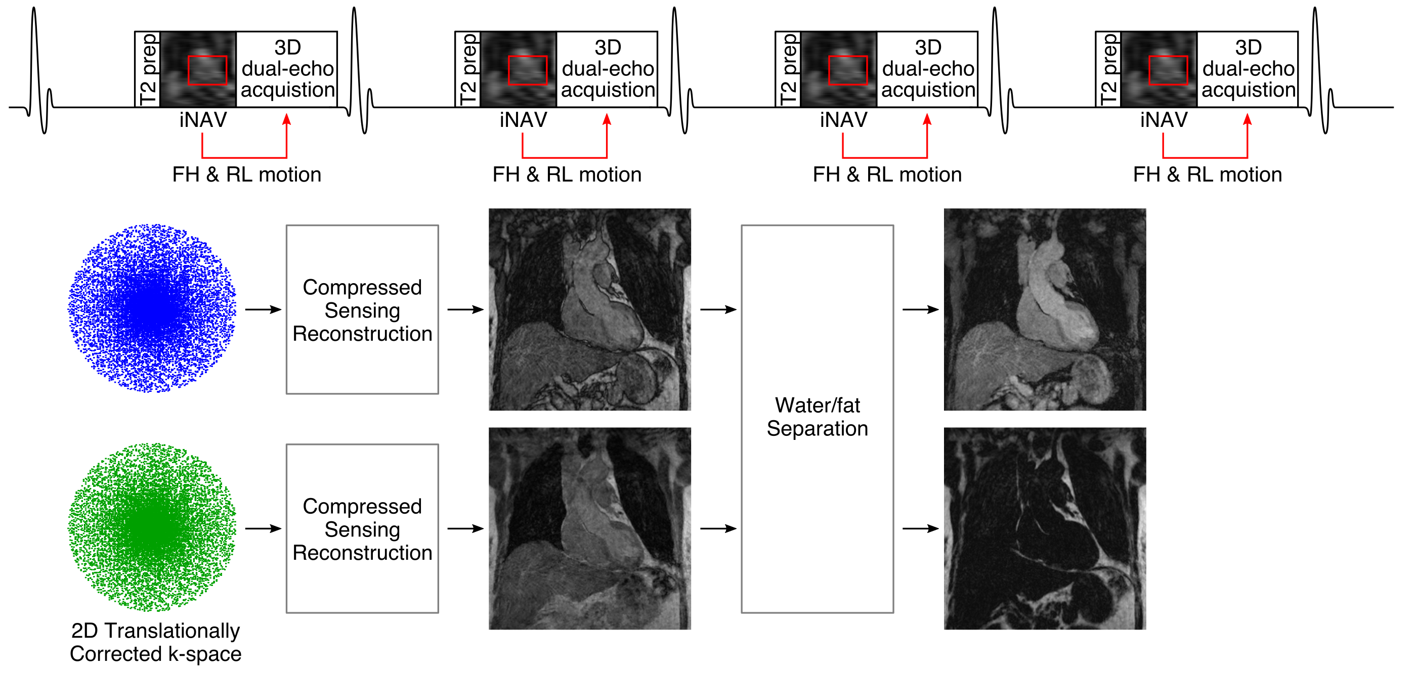
The undersampled 3D dual-echo CMRA dataacquisition sequence consists of an ECG-triggered spoiled gradient echo scheme following the VD-CAPSR trajectory, so that one spiral interleaf is acquired at each heartbeat (Fig 1). 2DiNAVs are acquired before the dual-echo 3D acquisition with low flip-angle excitation pulses, and are used to estimate and correct forfoot-head (FH) and right-left (RL) motion in a beat-to-beat fashion. The proposed acquisition and reconstruction approach was implemented as a prototype on a 3T scanner (MAGNETOM Skyra, Siemens Healthcare, Erlangen, Germany).
Seven subjects (46±17 years, 4 male) were scanned using the proposed dual-echo 3D CMRA sequence and the following imaging parameters: coronal orientation, resolution = 1 mm3 isotropic, FOV = 320×320×112-128mm3, TR/TE1/TE2 = 5.74/1.30/2.76ms, bipolar gradient readout, bandwidth = 965 Hz/px for both echoes, FA=15°, 4-fold undersampling. The motion-corrected, undersampled data were reconstructed using the FISTA algorithm and wavelet regularisation4. The optimization was run for 20 iterations and the regularization parameter was fixed to 0.001. A subject-specific trigger delay and acquisition window (90-120ms) were set coinciding with the mid-diastolic rest period. In four of the subjects, an additional SPAIR fat-suppressed CMRA acquisition with matching imaging parameters and undersampling factor was performed for comparison purposes.
Results
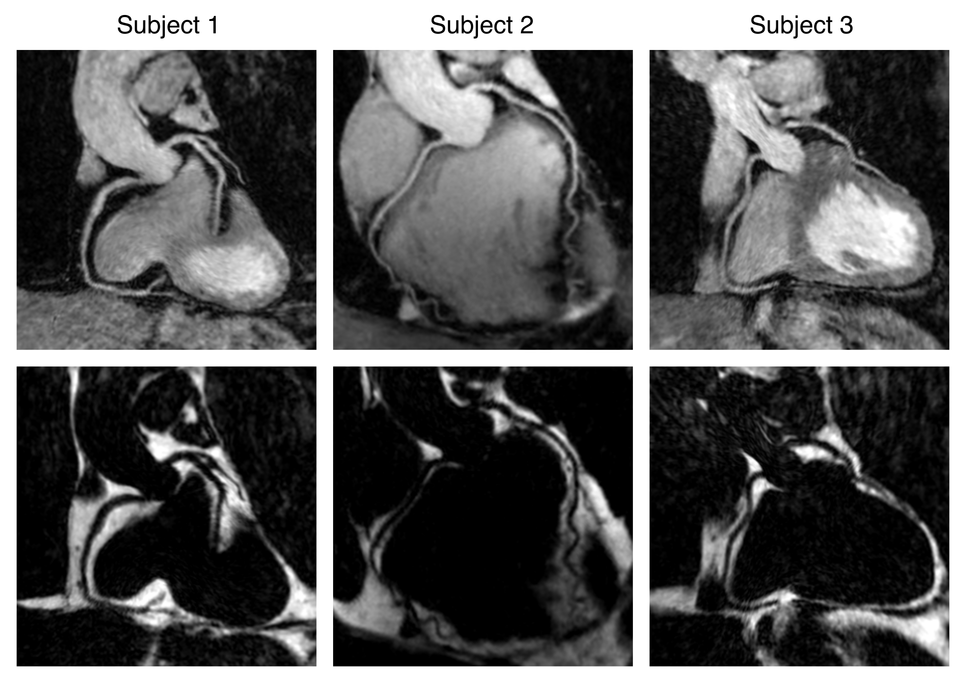
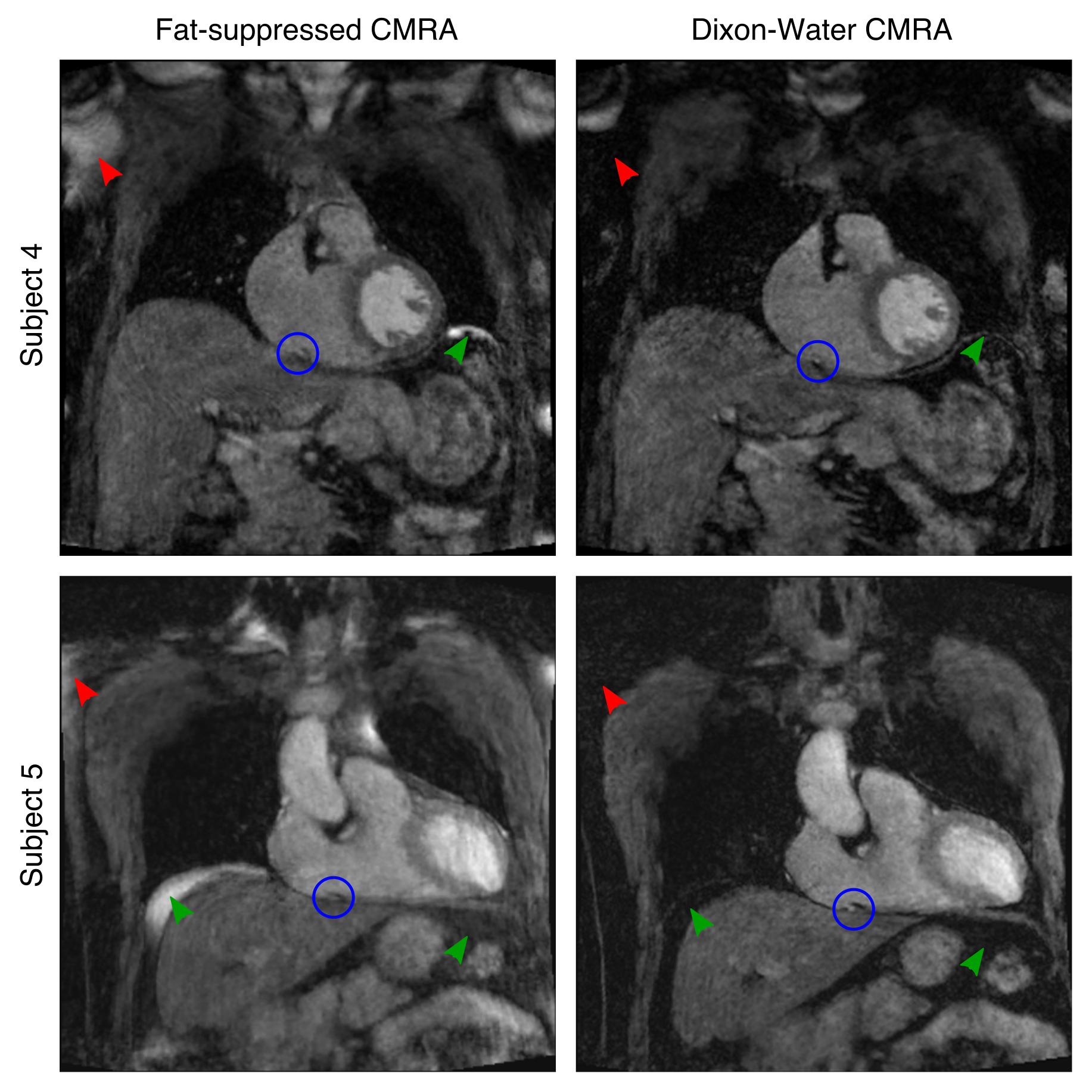
Water/fat CMRA scans were successfully completed in all subjects, with an average scan time of 9.83±1.45 min. Water/fat CMRA images and fat-suppressed CMRA images were reformatted to visualise simultaneously the right (RCA) and left (LAD) coronary arteries. The high spatial resolution of 1mm3 enables visualisation of both arteries in the proximal and distal segments in the water images, while also providing a complementary image with good depiction of cardiac fat (Fig 2). Compared to conventional fat-suppressed CMRA, improvements in suppression of subcutaneous (Fig 3, red arrows) and visceral fat (Fig 3, green arrows) can be observed, resulting in improved depiction of the coronary arteries (Fig 3, blue circles). The proposed water/fat approach produces a complementary high-resolution fat image (Fig 4) that can potentially be used for diagnostic purposes.Conclusion
Here we present an efficient framework for high-resolution whole-heart water/fat 3D CMRA imaging. This framework integrates image navigation to enable 2D translational respiratory motion correction, 100% respiratory scan efficiency and predictable scan time, and an undersampled acquisition and reconstruction framework for imaging in ~10 min. This framework results in high-resolution motion-corrected images with excellent fat suppression and diagnostic image quality, enabling the visualisation of the coronary anatomy and providing a complementary image of cardiac fat. The acquisition and undersampled translational motion-corrected reconstruction framework were fully implemented on a 3T scanner, thereby facilitating clinical translation.Acknowledgements
This work was supported by EPSRC (EP/L015226/1, EP/P001009, EP/P007619, EP/P032311/1) and Wellcome EPSRC Centre for Medical Engineering (NS/A000049/1).References
1. Nezafat M, Henningsson M, Ripley DP, et al. Coronary MR angiography at 3T: fat suppression versus water-fat separation. Magn Reson Mater Physics, Biol Med 2016;29:733–738
2. Bustin A, Ginami G, Cruz G, et al. Five-minute whole-heart coronary MRA with sub-millimeter isotropic resolution, 100% respiratory scan efficiency, and 3D-PROST reconstruction. Magn Reson Med 2018 doi: 10.1002/mrm.27354.
3. Liu J, Rapin J, Chang T, et al. Dynamic cardiac MRI reconstruction with weighted redundant Haar wavelets. In: Proceedings of the 20th Annual Meeting of ISMRM, Melbourne, Australia. 2012. p. 4249.
4. Forman C, Tillmanns C, Zenge MO, Schmidt M. Initial Experience in Patients for Highly Accelerated Free-Breathing Whole-Heart Coronary MRA. In: Proceedings of the 22th Annual Meeting of ISMRM, Milan, Italy. 2014, p. 0179.
Figures