1252
Pushing fMRI spatial and temporal resolution further: high density receive arrays combined with shot-selective 2D CAIPIRINHA for 3D EPI scans at 7T1Department of Radiology, University Medical Center Utrecht, Utrecht, Netherlands, 2Department of Radiotherapy, University Medical Center Utrecht, Utrecht, Netherlands, 3Department of Neuroscience, University of Turin, Turin, Italy, 4Philips Healthcare, Best, Netherlands
Synopsis
There is an overall drive to high resolution brain MRI with a short scan time. In this study, it was investigated whether 3D EPI fMRI scans acquired with high density receive arrays, can still benefit from a CAIPI sampling pattern, in terms of (temporal) SNR. A 2D CAIPIRINHA acceleration scheme for multi-shot 3D EPI scans was implemented. When combining this implementation with high density receive coil arrays at 7T, it allowed substantial reduction of the scan time for sub-millimeter fMRI scans of the visual cortex.
Introduction
To examine dynamic and spatially detailed brain functions using functional MRI (fMRI), the combination of both a high temporal resolution and a high spatial resolution is essential. Nevertheless, fMRI with both a high temporal resolution (<1 sec) and a high spatial resolution (<1 mm) combined, is rarely seen. In pursuit of high resolution scanning, the fMRI field shifts towards measurements with high density surface coil receive arrays that have many small coils closely packed together. These receive arrays facilitate the use of high acceleration factors with reduced g-factors at high resolutions [1-3]. Alternatively, parallel imaging techniques such as 2D CAIPIRINHA (CAIPI) [4], prove to be effective in reducing scan time of 3D EPI sequences as well [5-8].
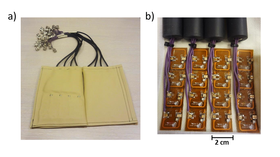
Since both high density receive arrays and CAIPI techniques aim to reduce scan time by enhancing the variation in spatial sensitivity, it can be questioned whether these techniques complement or compete with each other. Can a combination of these two approaches enable sub-mm and sub-sec fMRI? Here we investigate whether sub-mm multi-shot 3D EPI fMRI scans acquired with high density receive arrays, still benefit from a CAIPI sampling pattern, in terms of (temporal) SNR and noise reduction (g-factor). A shot selective 2D CAIPI sequence was implemented for multi-shot 3D EPI scans, which, instead of adding extra gradients, leaves them out. The sequence was evaluated in combination with high density receive arrays (Figure 1) for sub-millimeter fMRI acquisitions at high acceleration.
Sequence implementation
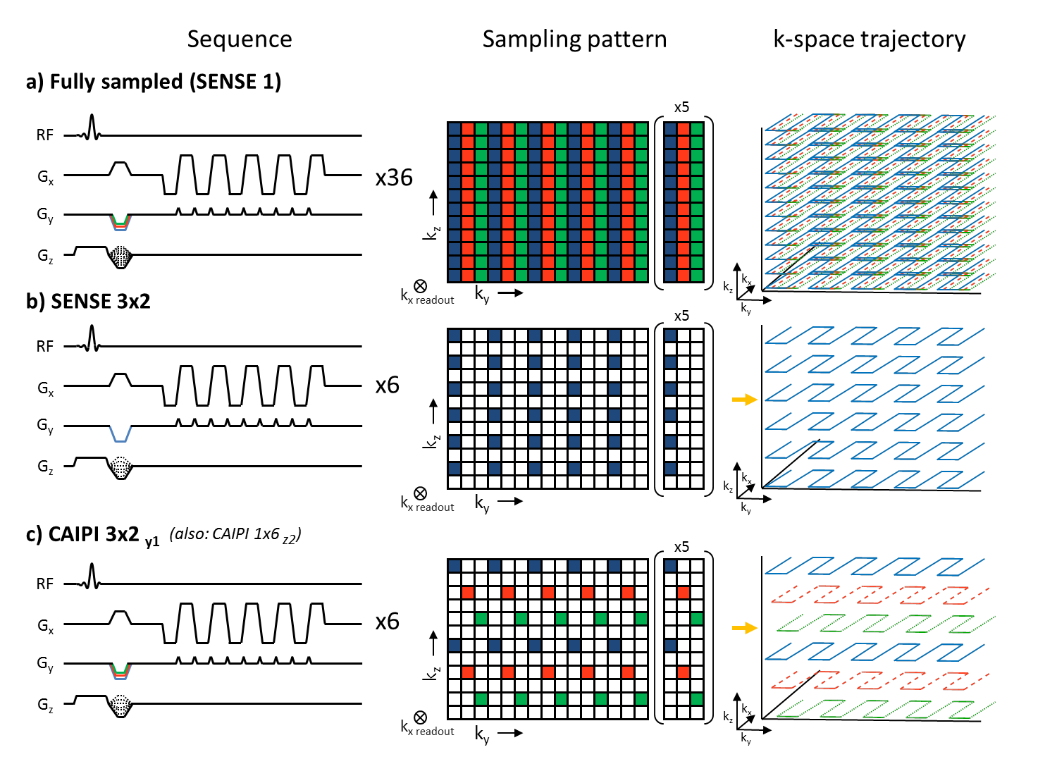
Several EPI shots of a multi-shot interleaved 3D EPI sequence were selectively skipped resulting in a CAIPI sampling pattern (Figure 2).Methods
Two participants were scanned in a 7 Tesla Achieva system (Philips, Best, the Netherlands). A birdcage volume coil for the head was used as transmit setup (Nova Medical, USA). The receive setup (Figure 1) consisted of 2x16 channel high density surface coil receive arrays [1] (MR Coils BV, Zaltbommel, the Netherlands).
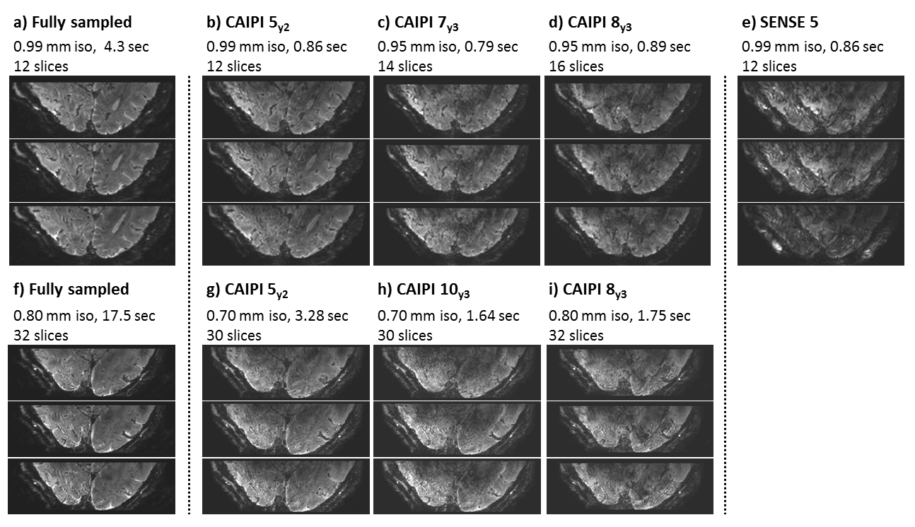
- Multiple 3D EPI scans of the visual cortex were acquired at rest, with different values for FOV, spatial and temporal resolution. Scan parameters are TR/TE= 54/27 ms, 20° flip angle, a resolution of 0.7-0.99 mm3 isotropic, a FOV ranging from 50x164x21 mm3 to 50x175x26 mm3, 12-30 slices, EPI factor 23-33, factor 5-10 CAIPI/SENSE acceleration, 0.79-3.28 sec dynamic scan time.
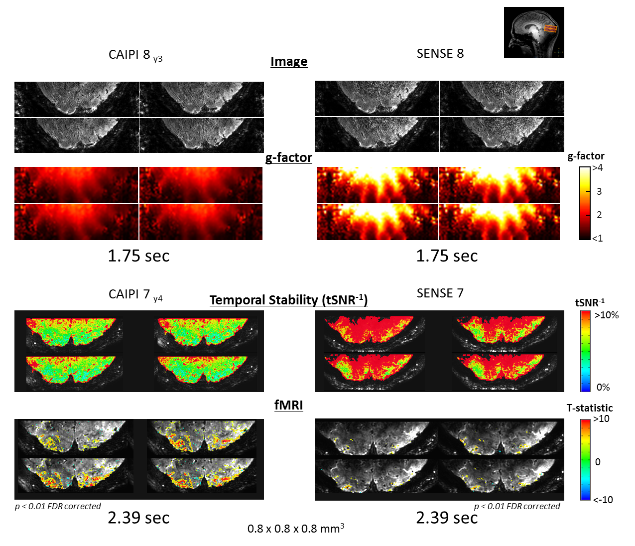
- fMRI data of the visual cortex was acquired during rest and during visual stimulation. Scan parameters fMRI: 3D EPI, TR/TE= 54/28 ms, 20° flip angle, 0.8x0.8x0.8mm3 voxels, 51x162.5x28mm3 FOV, 35 slices, matrix size 64x203, EPI factor 29, CAIPI factor 7 shift 4, volume acquisition 2.39 sec, a total scan time of 4 minutes. An identical scan was made with SENSE 7, as a reference. The visual stimulus consisted of a checkerboard that reversed contrast at 8 Hz.
All scans are reconstructed offline in a modified Philips Recon2.0 environment.
Results
Sub-millimeter 3D EPI scans acquired with high density receive arrays and different combinations for scan time and resolution are displayed in Figure 3. As the resolution improves, the scan time increases as well. Note that it is possible to perform sub-millimeter and sub-second EPI imaging of the visual cortex, without losing much image quality. The fMRI scans are displayed in Figure 4, both for CAIPI and SENSE undersampling. The figure shows fMRI scans, g-factor maps and tSNR maps. The spatial extent of fMRI activation is larger for the CAIPI scans. This is in line with the lower noise amplification (g-factor) and higher temporal SNR (improved stability 1/tSNR %) for the CAIPI scans at rest.Discussion and conclusion
The combination of high density receive coils and shot selective 2D CAIPI for 3D EPI, pushes the temporal resolution of sub-mm fMRI scans. The temporal SNR of the CAIPI scans is higher than the SENSE scans, also when using high density receive arrays. This gain in temporal SNR demonstrates that the benefit of CAIPI with the high dense arrays can be used to complement both techniques for maximizing spatial and temporal resolution. The sequence employed here is practical to implement, since it does not require SAR-demanding (multiband) RF pulses or extra gradient blips as compared to a multi-shot 3D EPI sequence. Furthermore, most EPI phase correction can be left in place. Overall, high density receive arrays in combination with shot selective 2D CAIPI for 3D EPI proves to be a valuable tool for reducing scan time of sub-millimeter fMRI acquisitions.
Acknowledgements
This work was supported by the Netherlands Organization for Scientific Research (NWO), specifically research grants: 040.11.581, ALW-834.14.004 and Vidi Grant 13339 (Petridou).References
[1] N. Petridou et al. (2013) Pushing the limits of high-resolution functional MRI using a simple high-density multi-element coil design. NMR Biomed 26: 65–73
[2] D.A. Feinberg et al. (2018) Pushing the limits of ultra-high resolution human brain imaging with SMS-EPI demonstrated for columnar level fMRI. Neuroimage 164:155-163
[3] A. Fracasso et al. (2018) Laminar imaging of positive and negative BOLD in human visual cortex at 7T. Neuroimage 164:100-111
[4] F.A. Breuer et al. (2006) Controlled Aliasing in Volumetric Parallel Imaging (2D CAIPIRINHA). Magn Reson Med 55:549–556
[5] B.A. Poser et al. (2014) Accelerated 3D EPI using 2D blipped-CAIPI for high temporal and/or spatial resolution. Proceedings of the 22th Annual Meeting of the ISMRM Milan. pg.#1506.
[6] M. Narsude et al. (2016) Three-Dimensional Echo Planar Imaging with Controlled Aliasing: A Sequence for High Temporal Resolution Functional MRI. Magn Reson Med 75:2350–2361
[7] F.A. Breuer et al. (2008) Zigzag Sampling for Improved Parallel Imaging. Magn Reson Med 60:474–478
[8] W. Zwaag et al. (2018) High Spatio-Temporal Resolution in Functional MRI With 3D Echo Planar Imaging Using Cylindrical Excitation and a CAIPIRINHA Undersampling Pattern. Magn Reson Med 79(5):2589-2596
Figures