1245
Differentiation of Malignant and Benign Parotid Tumors with Amide Proton Transfer-Weighted MR Imaging1Peking Union Medical College Hospital, Beijing, China, 2Philips Healthcare, Beijing, China
Synopsis
This retrospective study evaluated the diagnostic efficacy of diffusion-weighted imaging (DWI) and amide proton transfer-weighted (APTw), for differentiating between malignant, pleomophic adenomas and Warthin’s tumours. APTw imaging can differentiate Warthin’s tumors and malignant tumors of parotid. ADC can differentiate Warthin’s tumors and pleomorphic adenomas. APT combined with ADC had the better performance to differentiate the three common kinds of parotid tumours.
Introduction/Purpose:
Modern imaging plays a key role in discriminating benign and malignant parotid tumours. Over the past decades, the roles of functional MRI, such as diffusion-weighted imaging (DWI) have been evaluated in head and neck lesions.DWI quantifies the diffusion mobility of water protons with apparent diffusion coefficient (ADC).[1] However, additional ability of functional MRI in differential diagnosis of parotid lesions is still controversial because overlap is detected between malignant and benign parotid tumours.[2] Amide proton transfer-weighted (APTw) MR imaging based on the chemical exchange saturation transfer (CEST) mechanism, can detect mobile proteins and peptides in tissues, without an exogenous contrast agent.[3] The aim of our study is to investigate the ability of APTw imaging, in comparison with that of diffusion-weighted imaging (DWI), to differentiate malignant and benign parotid lesions.Methods and materials:
A retrospective review of patients who underwent MR examination including APTw imaging and DWI for evaluation of parotid lesions were enrolled, between October 2017 and September 2018. The patients were divided into malignant, pleomophic adenomas and Warthin’s tumours, based on pathological results. APTw imaging was performed on a clinical whole-body 3.0-Testla MR system (Ingenia CX 3.0T, Philips Healthcare, Best, the Netherlands). APT signal intensity (SI) was defined as the asymmetry magnetization transfer ratio at 3.5 ppm and was mapped. An apparent diffusion coefficient (ADC) map was generated using b-values of 0 and 800 s/mm2. APT SI and ADC were calculated by placing regions-of-interest in the tumours on these maps. One-way ANOVA analysis was used to compare the difference between different pathological results. Receiver operating characteristic (ROC) analysis was performed and the area under the ROC curve was calculated for ADC value and APT SI that were significantly different (P<0.05) . Sensitivity (Se), specificity (Sp), positive predictive value (PPV), negative predictive value (NPV), and accuracy were calculated using the cut-off value with the highest AUC.Results:
A total of 18 patients (10 males and 8 females; age: 56.72±13.20 years) were included, consisting of 6 malignant tumours, 7 pleomorphic adenomas and 5 Warthin’s tumours. Malignant tumours included 2 mucoepidermoid carcinoma, 1 metastasis carcinoma, 1 low-grade adenocarcinoma, 1 adenoid cystic carcinoma and 1 acinar cell carcinoma. Except 1 Warthin’s tumour and 1 pleomorphic adenoma, 16 patients received DWI scanner. The mean ADC value of Warthin’s tumours (0.747×10⁻³mm2S-1) was significantly lower than that of pleomorphic adenomas (1.813×10⁻³mm2S-1, P=0.01). However, there was no significant difference between malignant tumours (1.295×10⁻³mm2S-1) and pleomorphic adenomas (P=0.058). AUC of mean ADC to identify Warthin’s tumours was 0.958±0.051(P=0.008). The cutoff value of mean ADC was 1.035 (Youden indexmax=0.833), and corresponding diagnostic performance were: Se=100% (4/4), Sp=83.3% (10/12), PPV=66.7% (4/6), NPV=100% (10/10), and accuracy=87.5% (14/16).Except 2 malignant tumours and 2 pleomorphic adenomas with significant cystic change, 14 patients went to APT analysis. The mean APT SI of malignant tumours (3.72±2.30%) was significantly higher than that of Warthin’s tumours (1.14±1.61%,P=0.049). However, there was no significant difference between malignant tumours and pleomorphic adenomas (2.14±1.75%, P=0.381). AUC of mean ADC to identify malignant tumours was 0.875±0.096(P=0.034). The cutoff value of mean ADC was 3.165 (Youden indexmax=0.8), and corresponding diagnostic performance were: Se=100% (4/4), Sp=80% (8/10), PPV=66.7% (4/6), NPV=100% (8/8), and accuracy=87.5% (12/14).Conclusion:
Malignant tumours had the highest APT SI and Warthin’s tumours had the lowest ADC value. APT combined with ADC could differentiate the malignant, pleomorphic adenomas and Warthin’s tumours of parotid.Acknowledgements
No acknowledgement found.References
[1] Lee YY, Wong KT, King AD, Ahuja AT. Imaging of salivary gland tumours. Eur J Radiol. 2008. 66(3): 419-36.
[2] Yuan Y, Tang W, Tao X. Parotid gland lesions: separate and combined diagnostic value of conventional MRI, diffusion-weighted imaging and dynamic contrast-enhanced MRI. Br J Radiol. 2016. 89(1060): 20150912.
[3] Yu L, Li C, Luo X, et al. Differentiation of Malignant and Benign Head and Neck Tumors with Amide Proton Transfer-Weighted MR Imaging. Mol Imaging Biol. 2018 .
Figures
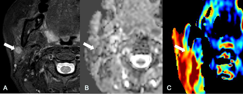
Fig 1. A 49-year-old man was found a tumour in left parotid. The T2 weighted imaging with fat suppression (A) showed a lesion with high signal intensity in the left parotid. The mean ADC value (B) was 0.98×10⁻³mm2S-1.The mean APT SI (C) was 1.77%. The pathological result was Wathin's tumour.