1224
Contrast-enhanced MRI reveals differential effects of afferent and efferent vagus nerve stimulation on gastric motility1Electrical and Computer Engineering, Purdue University, West Lafayette, IN, United States, 2Biomedical Engineering, Purdue University, West Lafayette, IN, United States, 3Psychological Sciences, Purdue University, West Lafayette, IN, United States
Synopsis
We used in vivo contrast-enhanced MRI to evaluate how vagus nerve stimulation (VNS) could modulate and coordinate the motility of the antrum and pylorus. We tested cervical VNS with different settings (afferent or efferent) and parameters (amplitude, duration, and frequency) and found differential effects on the motility. Our results suggest that selective stimulation of vagal afferent using a monophasic pulse train is a more effective strategy to facilitate and coordinate the antro-pyloric motility than selective stimulation of the vagal efferent. These results could shed lights on the selection of VNS settings for modulating gastric functions.
Purpose
Vagus nerve stimulation (VNS) is an emerging bioelectronic therapy to remedy gastric disorders [1]. However, the efficacy of different VNS parameters on modulating gastric functions remains unclear. Moreover, concurrent VNS settings have only focused on activating the motor limb of the brain-gut circuitry (e.g. by stimulating efferent vagal fibers), leaving the therapeutic potential of afferent stimulation that engages the complete reflex arcs undescribed. Here, we set out to disentangle the effects of graded VNS settings and directionality preferences of VNS on gastric functions.Method
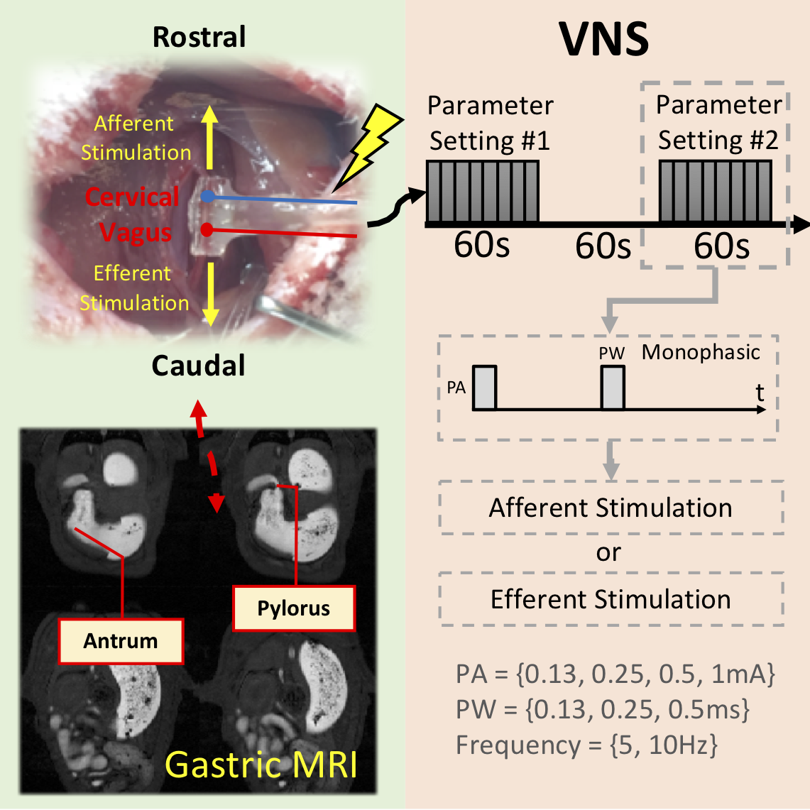
Thirteen Sprague-Dawley rats (male, 266-338g) were studied according to our protocol approved by the Purdue Animal Care and Use Committee. The rats were trained to voluntarily consume ~10g of gadolinium(Gd)-labeled meal after an 18-hour fast. Soon after meal ingestion, the animals were anesthetized with 4% Isoflurane for 5 minutes, followed by implantation of a bipolar cuff electrode on the left cervical vagus nerve. Prior to imaging, they first received a subcutaneous (SC) bolus injection of 0.01mg/kg dexmedetomidine solution (0.05mg/ml, Zoetis), followed by a continuous SC infusion of dexmedetomidine at 0.03mg/kg/hr along with 0.3-0.5% isoflurane mixed with oxygen at a flow rate of 800ml/min 15 min after the bolus. The animals were then scanned in a 7T small-animal MRI system (BioSpec 70/30, Bruker). A respiratory-triggered FLASH sequence was used to obtain T1-weighted abdominal images (TR/TE = 11.8/1.1ms; FA = 25°; matrix size = 128 x 128; slice thickness = 1.5mm; FOV = 60 x 60mm; and 4 slices). Readout gradient triggers were recorded to adjust acquisition times between volumes due to varying respiratory rates. After acquiring 4 minutes of baseline motility images, VNS was delivered simultaneously with MRI acquisition. Of the 13 animals, 8 were imaged under afferent VNS (i.e. action potential propagates rostrally) and 5 were imaged under efferent VNS (i.e. action potential propagates caudally). The direction in which action potential traveled was controlled by the placement of the cathode on the bipolar electrode as shown in Fig. 1. Directionality preferences were confirmed by electrophysiological recordings in the nucleus tractus solitarius (NTS). For both afferent and efferent VNS, the parameters were varied in terms of pulse amplitude, width, and frequency. Each VNS setting comprised of a duty cycle of 1 minute on and 1 minute off, and different VNS settings were performed in a randomized order. Abdominal MRI images were processed using our previously developed software [2], where the amplitude of antral contraction and the degree of pyloric opening were quantified and compared against different VNS settings. The coordination between antral peristalsis and pyloric activity was further evaluated by computing the phase-locking value between the contraction time series at the antrum and the pylorus. All quantities measured under VNS were normalized against their baseline values within each animal and expressed as percentage change from baseline.Results
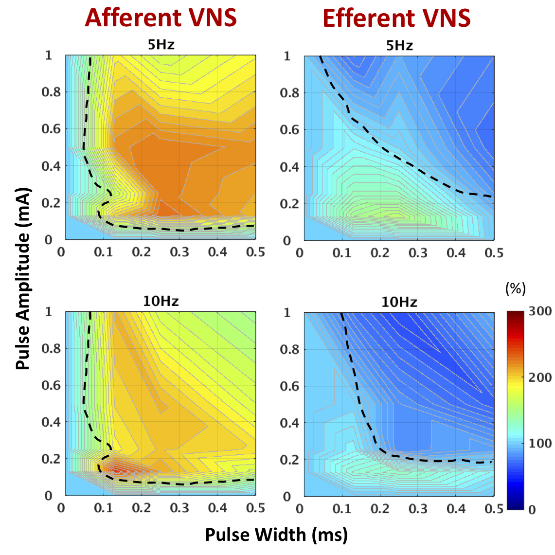
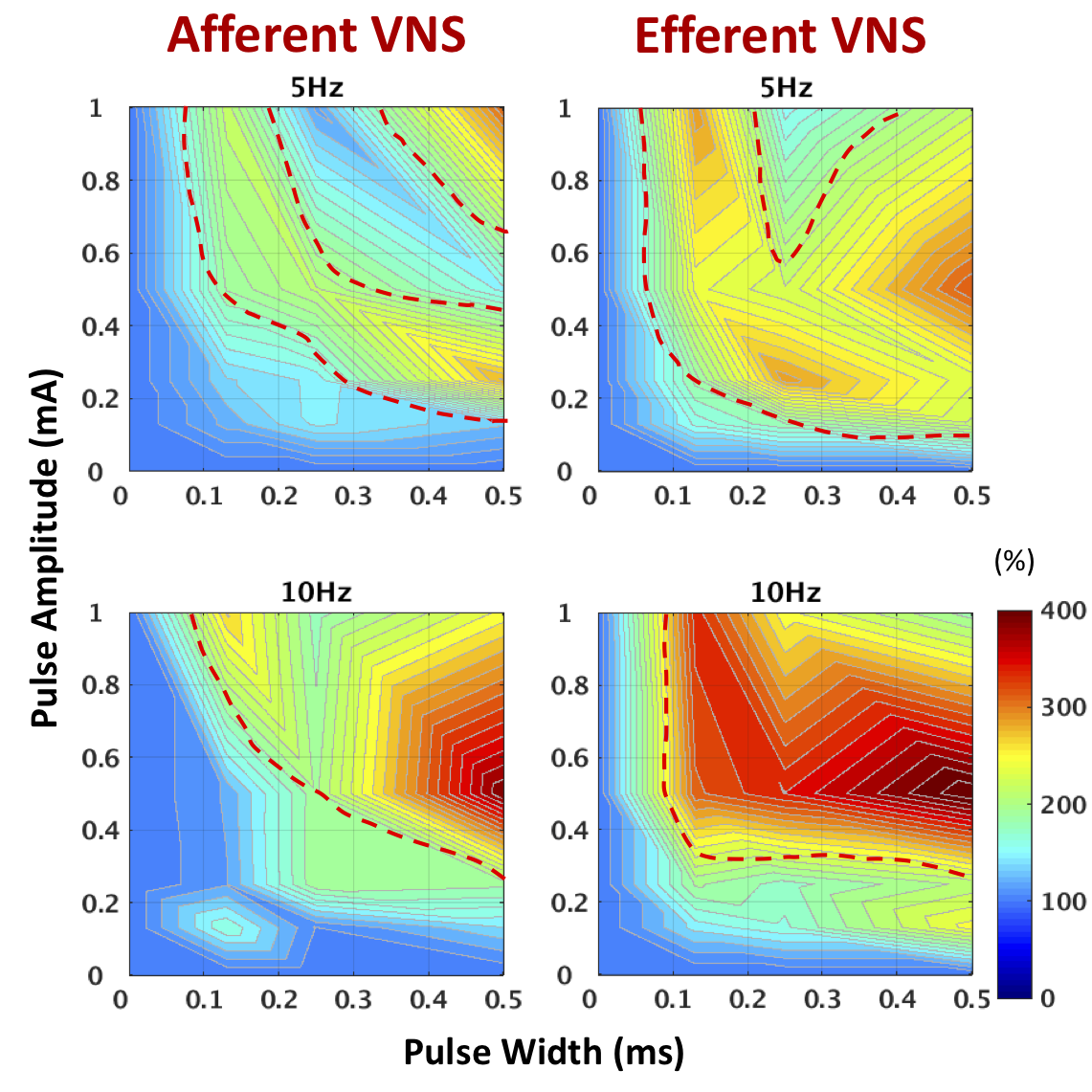
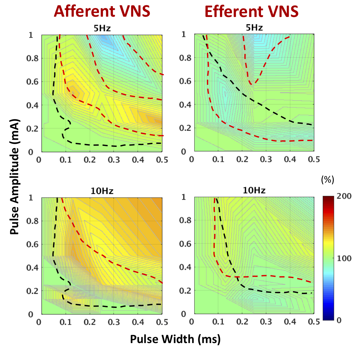
We observed that brainstem neuronal responses to electrical stimulation at the left cervical vagus was dependent on the polarity of the applied stimulation pulse. The neural responses at NTS was significantly larger when the cathode was placed cranial to the cuff, suggesting that monophasic VNS was able to activate afferent (or efferent) signal flow with reasonable selectivity. With our gastric MRI protocol and analysis, we found that afferent VNS induced stronger antral contraction than efferent stimulation almost under all tested VNS settings (Fig. 2). In particular, efferent VNS dampened antral contraction and induced excessive secretion in the antrum when applied at higher doses. Interestingly, both efferent and afferent VNS promoted pyloric opening, perhaps with the former being more effective under a wider spectrum of VNS settings (Fig. 3). Lastly, we found that only afferent VNS could promote antro-pyloric coordination while enhancing their motility (Fig. 4), suggesting that activating the reflex arc of the vagovagal circuitry is potentially a more favorable approach to modulate gastric functions.Conclusion
Here, we report the use of contrast-enhanced MRI to investigate a possible differential effect of afferent versus efferent cervical VNS on gastric motility under a range of stimulation parameters in rodents.Gastric MRI data revealed that electrical activation of the afferent pathway may promote gastric motility and coordination more effectively then directly activating the efferent pathway. A reduction in antral contraction amplitude and relaxation of pyloric sphincter under efferent VNS highlighted the inhibitory pathway of the motor limb of the vagovagal circuitry.The physiology data also indicated that efferent stimulation was often accompanied with undesirable off-target effects such as bradycardia and bradypnea, which were in line with the observations from previous VNS studies.Acknowledgements
This work was supported by NIH SPARC 1OT2TR001965.References
[1] Lu K-H, et al., “Vagus nerve stimulation promotes gastric emptying by increasing pyloric opening measured with magnetic resonance imaging,” Neurogastroenterology and Motility, 30(10): e13380, 2018.
[2] Lu K-H, et al., "Contrast enhanced magnetic resonance imaging of gastric emptying and motility in rats," IEEE Transactions on Biomedical Engineering. 2017;64(11):2546-2554.
Figures