1219
Quantitative 129Xe MRI detects early impairment of gas-exchange in a rat model of pulmonary arterial hypertension1Biomedical Engineering, Duke University, Durham, NC, United States, 2Radiology, Duke University Medical Center, Durham, NC, United States, 3Department of Medicine, Duke University Medical Center, Durham, NC, United States
Synopsis
Hyperpolarized 129Xe MRI is capable of regional mapping of gas-exchange and has found application in a wide range of lung disorders. Here, we apply 129Xe gas exchange MRI and dynamic spectroscopy at 7 Tesla to study a rat model of pulmonary arterial hypertension. 129Xe spectroscopy and gas-exchange imaging showed reduced uptake by RBCs early in the progression of the disease, and at later time points was accompanied with increased uptake by barrier tissues, structural abnormalities, edema, and ventilation defects. Imaging results were compared to H&E histology, which showed evidence of vascular remodeling.
Introduction
The ability to image 129Xe in the lung
airspaces, its uptake in interstitial barrier tissues and its transfer to red
blood cells (RBCs) enables direct probing of lung structure and function, and
the assessment of gas-exchange at the pulmonary-capillary level. In this study,
we used 3D quantitative hyperpolarized (HP) 129Xe gas-exchange MRI
and spectroscopy, combined with anatomical 1H MRI, to detect lung
function impairment in a rat model of pulmonary arterial hypertension (PAH).
Methods
The monocrotaline (MCT) model was selected for its ability to generate pulmonary lesions similar to human PAH1 in male Sprague-Dawley rats. Control animals (N=8) received no treatment, while the PAH group (N=9) received one injection of MCT (60 mg/kg subcutaneous). The cross-sectional study imaged treated rats at one of two possible time points post-injection of MCT: 1 week (N=4) and 2 weeks (N=5).
Prior to imaging in a 7-T preclinical magnet (BioSpec 70/20, Bruker, Billerica, MA, USA), animals were anesthetized, intubated and connected to an HP-gas compatible ventilator2. Imaging included anatomical 1H MRI, 129Xe ventilation and dissolved-phase MRI. 129Xe spectra were acquired every 20-ms over several breaths. Parameters for all acquisition are listed in Figure 1. 129Xe gas- and dissolved-phase images were processed to create maps of ventilation, barrier tissue-uptake and RBC-transfer3,4. Dynamic 129Xe spectroscopy was processed to quantify the magnitude of cardiogenic oscillations in the amplitude of the RBC resonance5.
Immediately after imaging, the rats were euthanized and their lungs extracted for histology6.
Results
Figure 2 shows H&E histology in a control and week-2 PAH rat. In the PAH rat, lung tissue exhibited mild thickening of the endothelial layer, and moderate smooth muscle proliferation.
Figure 3 shows the static and dynamically acquired spectra. Rats in the PAH group show diminished RBC:barrier signal. The RBC:barrier was 0.47±0.03 in the control group, and significantly reduced in the PAH group to 0.40±0.06 (p=0.014). The reduction in the RBC:barrier was observed as early as 1-week post MCT-injection.
Figure 3C shows 129Xe-RBC transfer amplitude variation averaged over 5 consecutive breaths. These oscillations were quantified by fitting to a sine function, and were identified to be of ~5 Hz frequency, consistent with the heart rate of the animals. The magnitude of cardiogenic oscillations appeared greater in the PAH group, although not statistically significant.
Gas exchange maps from control animals (Figure 4) exhibited homogeneous, normal ventilation. The barrier-uptake and RBC-transfer images showed signal predominantly in the normal range, but also exhibited baseline defects, mostly near the base of the lungs and below the heart.
In the PAH
group, ventilation remained largely normal, while 2 out of 4 PAH animals at week 1 exhibited regions of
elevated barrier uptake, and later, 4 out of 5 animals at week 2. Defects in
RBC-transfer were observed at both week 1 and 2, mostly confined to the
anterior lung (Figure 4B, red arrows).
RBC-transfer defect percentage was significantly higher in the PAH than control groups (17.5±5.2 % vs 12.6±3.8 %, p=0.049), with two PAH rats exhibiting striking defects comprising 26% of the lung (Figure 4 D).
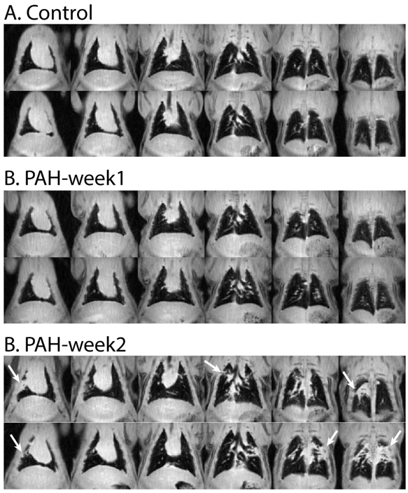
1H images revealed a normal thoracic cavity in the control group (Figure 5), whereas the PAH groups exhibited regions of edema (white arrows).
Discussion
In this rat model of PAH, impaired gas-exchange as reflected by 129Xe transfer to RBC cells was detected as early as one week post-injection. As the disease progresses, regions of increased barrier uptake and peripheral ventilation defects become apparent, and are accompanied with regions of edema on 1H MRI. The latter is not typical found in human PAH, but is consistent with the widespread pneumotoxicity of MCT, which includes interstitial and alveolar edema7,8.
Evidence of interstitial thickening in the PAH model could explain the larger magnitude of oscillations in the amplitude of the 129Xe RBC resonance. This finding is consistent with human idiopathic pulmonary fibrosis5, and is postulated to arise from the cardiac output being directed to a reduced pulmonary capillary volume.
Conclusion
We have presented the first application of quantitative 3D 129Xe gas-exchange MRI to the well-established MCT rat model of PAH. This proof-of-concept study demonstrated the sensitivity of 129Xe spectroscopy and imaging to detect several characteristics of PAH. Particularly interesting was a significant reduction in 129Xe transfer to RBCs, which was correlated in imaging and spectroscopy. This suggests the ability to observe early markers of gas-exchange impairment before other structural abnormalities manifested.Acknowledgements
NIH/NHLBI R01 HL105643, Burroughs Wellcome Career Award for Medical Scientists (Rajagopal)
References
[1] Gomez-Arroyo JG, Farkas L, Alhussaini AA, Farkas D, Kraskauskas D, Voelkel NF, Bogaard HJ. The monocrotaline model of pulmonary hypertension in perspective. American journal of physiology Lung cellular and molecular physiology 2012;302(4):L363-369.
[2] Virgincar RS, Dahlke J, Robertson SH, Morand N, Qi Y, Degan S, Driehuys B, Nouls JC. A portable ventilator with integrated physiologic monitoring for hyperpolarized 129Xe MRI in rodents. J Magn Reson 2018;295:63-71.
[3] Wang Z, Robertson SH, Wang J, He M, Virgincar RS, Schrank GM, Bier EA, Rajagopal S, Huang YC, O'Riordan TG. Quantitative Analysis of Hyperpolarized 129Xe Gas Transfer MRI. Medical Physics 2017;44(6):2475-2428.
[4] He M, Kaushik SS, Robertson SH, Freeman MS, Virgincar RS, McAdams HP, Driehuys B. Extending Semi-Automatic Ventilation Defect Analysis for Hyperpolarized 129Xe Ventilation MRI. Acad Radiol 2014.
[5] Dynamic Spectroscopy, Elly
[6] Ma Z, Mao L, Rajagopal S. Hemodynamic Characterization of Rodent Models of Pulmonary Arterial Hypertension. Journal of visualized experiments : JoVE 2016(110).
[7] Lee Y-S, Byun J, Kim J-A, Lee J-S, Kim KL, Suh Y-L, Kim J-M, Jang H-S, Lee J-Y, Shin I-S. Monocrotaline-induced pulmonary hypertension correlates with upregulation of connective tissue growth factor expression in the lung. Experimental & molecular medicine 2005;37(1):27.
[8] Kiss T, Kovacs K, Komocsi A, Tornyos A, Zalan P, Sumegi B, Gallyas F, Kovacs K. Novel Mechanisms of Sildenafil in Pulmonary Hypertension Involving Cytokines/Chemokines, MAP Kinases and Akt. PLoS ONE 2014;9(8):e104890.
Figures
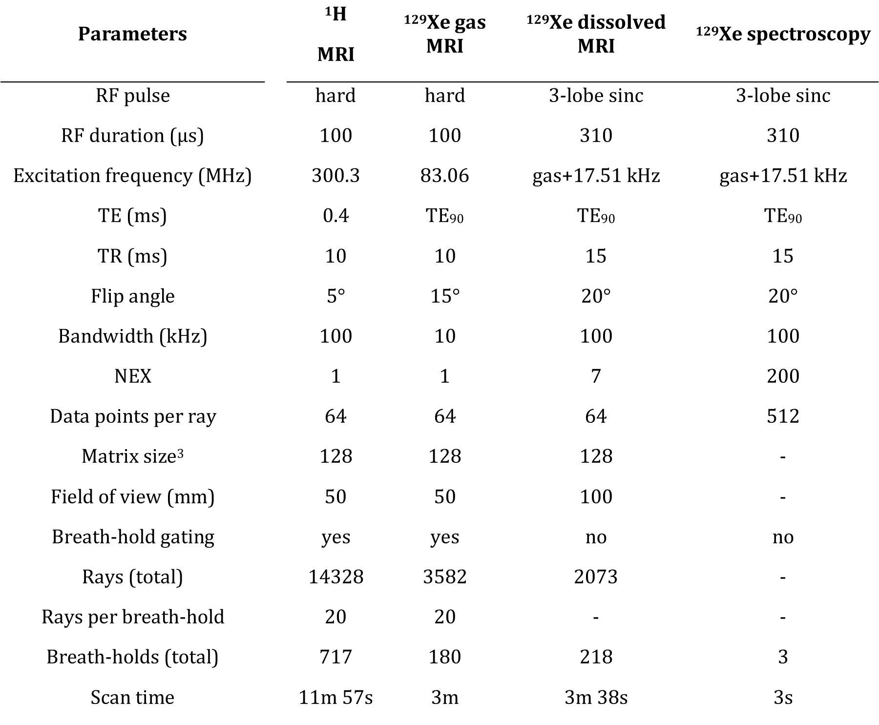
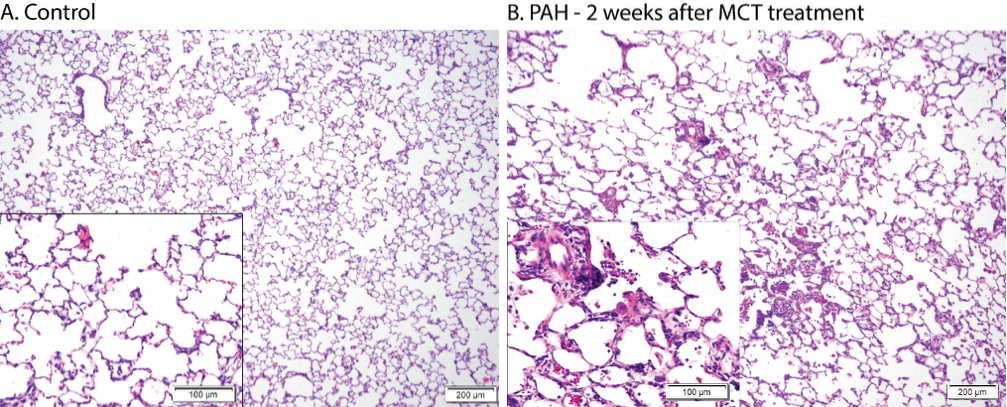
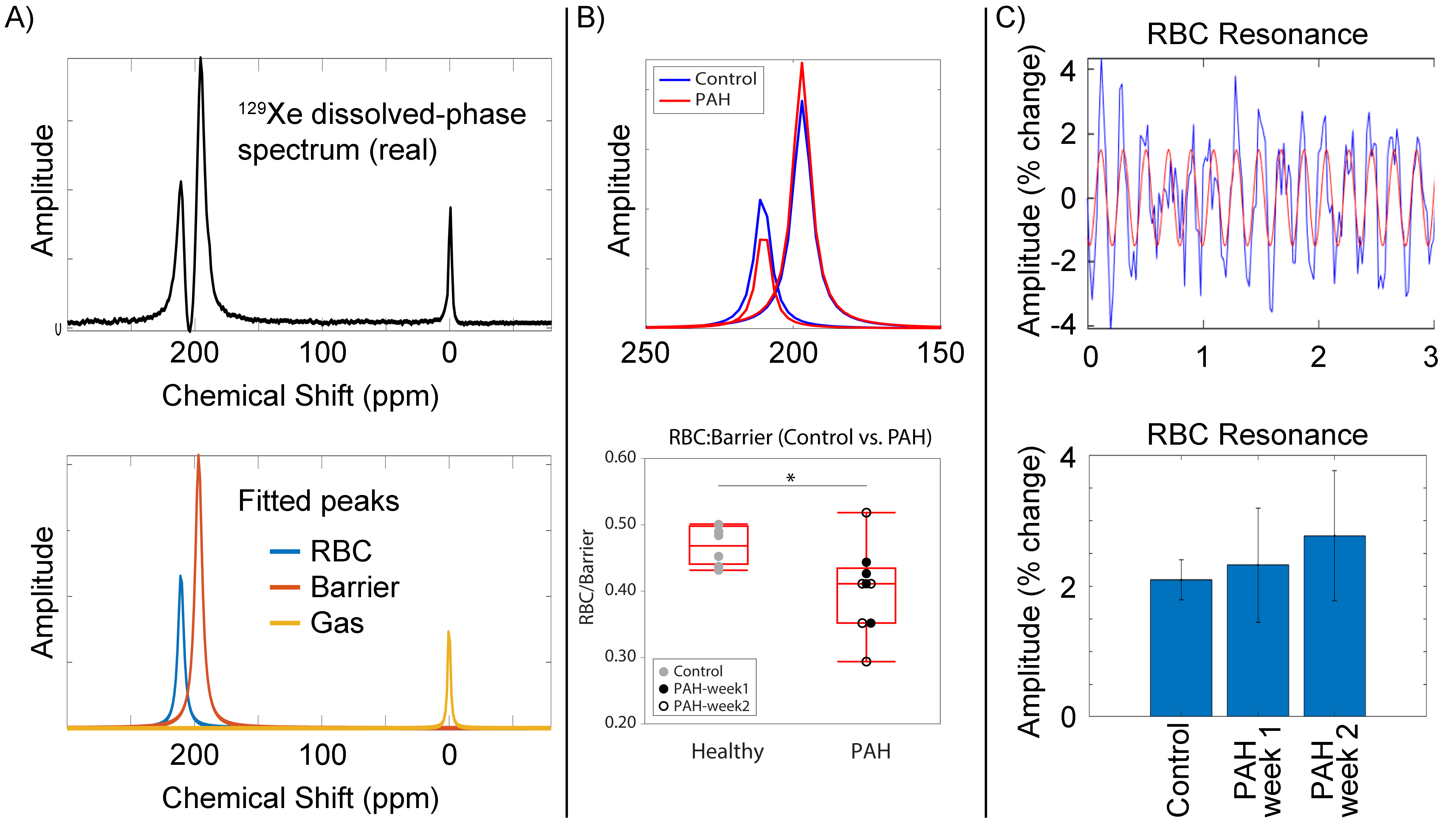
Figure 3: (A) 129Xe dissolved-phase spectrum of a healthy rat (top). The spectrum was fitted to extract the gas, barrier, and RBC resonances (bottom). (B) RBC and barrier resonances of a healthy rat compared to a PAH rat. In PAH rats, the ratio of the amplitudes of the RBC resonance vs barrier tissues (RBC:barrier) was found to be significantly reduced (p = 0.014). (C) variation of amplitude of the 129Xe RBC resonance over 3 consecutive breaths (blue). A fitted sine wave (red) was used to estimate the amplitude of oscillations, given by a bar plot (bottom).
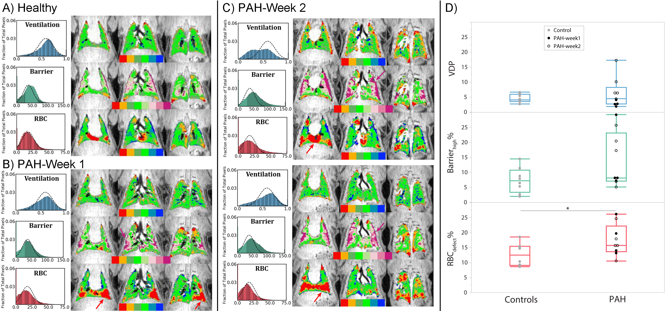
Figure 4: (A) Ventilation and gas-exchange maps with histograms in a healthy rat. The maps show homogeneous ventilation, barrier uptake, and RBC-transfer signal with a few baseline defects in barrier and RBC signal. (B-C) Maps in PAH rats. The prevalence of elevated barrier-uptake (purple arrows) increased from 50% of the animals at week 1, to 80% of the animals at week 2. Defects in RBC-transfer were observed in the PAH group at week 1 and 2 (red arrows). (D) Box-and-whisker plots for VDP, Barrierhigh, and RBCdefect for the control and PAH groups. RBCdefect was significantly higher in PAH.