1218
Click Your Fat Away: Rapid Synthetic Knee MR Imaging with Switchable Fat and Magnetization-Transfer Contrast1Advanced Clinical Imaging Technology, Siemens Healthcare AG, Lausanne, Switzerland, 2Department of Radiology, University Hospital (CHUV), Lausanne, Switzerland, 3LTS5, École Polytechnique Fédérale de Lausanne, Lausanne, Switzerland, 4Department of Radiology and Radiological Science, Johns Hopkins University School of Medicine, Baltimore, MD, United States, 5Radboud University Nijmegen, Donders Institute for Brain, Cognition and Behaviour, Nijmegen, Netherlands
Synopsis
Synthetic MRI is increasingly used in clinical practice to supplement or replace conventional MRI, due to the ability to generate both a multitude of MR images with weighted contrasts and quantitative T1, PD and T2 maps, which is of high interest in musculoskeletal imaging. To further broaden the application of synthetic image contrasts, we developed a method which allows switching between images with and without fat signal as well as magnetization-transfer weighting. The method includes three pulse sequences to estimate quantitative maps for the calculation of a variety of contrasts and requires a total acquisition time of 6:46 min.
Introduction
Quantitative MRI (qMRI) is a well-established technique in musculoskeletal (MSK) research. With the introduction of new acceleration techniques, qMRI is now becoming increasingly feasible for clinical practice. In addition to quantification, qMRI also allows for generating an arbitrary number of synthetic MR image contrasts. As the number of required qMRI pulse sequences can now be acquired in the same time or faster than individual conventional pulse sequences, qMRI has the potential to replace conventional pulse sequences. The use of synthetic images derived from a single T2-mapping sequence was shown to reduce acquisition times compared to individual acquisitions of morphological and quantitative sequences1. The addition of a T1 map allows for the incorporation of T1-related effects (e.g., short repetition time or inversion recovery STIR) and yields clinically useful MR images2. However, fat suppression has been limited to synthetic inversion recovery.
In this work, we describe a method which allows the additional features of “switching” the fat signal and the magnetization-transfer-weighting (MT-weighting) in synthetic contrasts “on” or “off”, which is a functionality that is expected to further benefit clinical MSK applications.
Methods
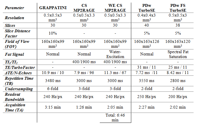
All acquisitions were performed at 1.5T (MAGNETOM Aera, Siemens Healthcare, Erlangen, Germany) using a 15-channel knee coil. After internal review board approval and written informed consent, four patients with knee pain were included. A T1 map was acquired using a compressed sensing-(CS)-accelerated prototype MP2RAGE acquisition3,4. A proton-density-weighted image M0P was also obtained by estimating the initial magnetization of this MP2RAGE sequence. The same CS-MP2RAGE sequence was repeated using a water-selective excitation (WE), in order to derive an initial magnetization image without fat signal M0W. In addition, a T2 map was acquired with a prototype GRAPPATINI5 sequence. The GRAPPATINI sequence also estimates its initial magnetization M0M which is magnetization-transfer weighted since it uses interleaved slice sampling6. The total acquisition time of the three sequences was 6:46 min (Table 1).
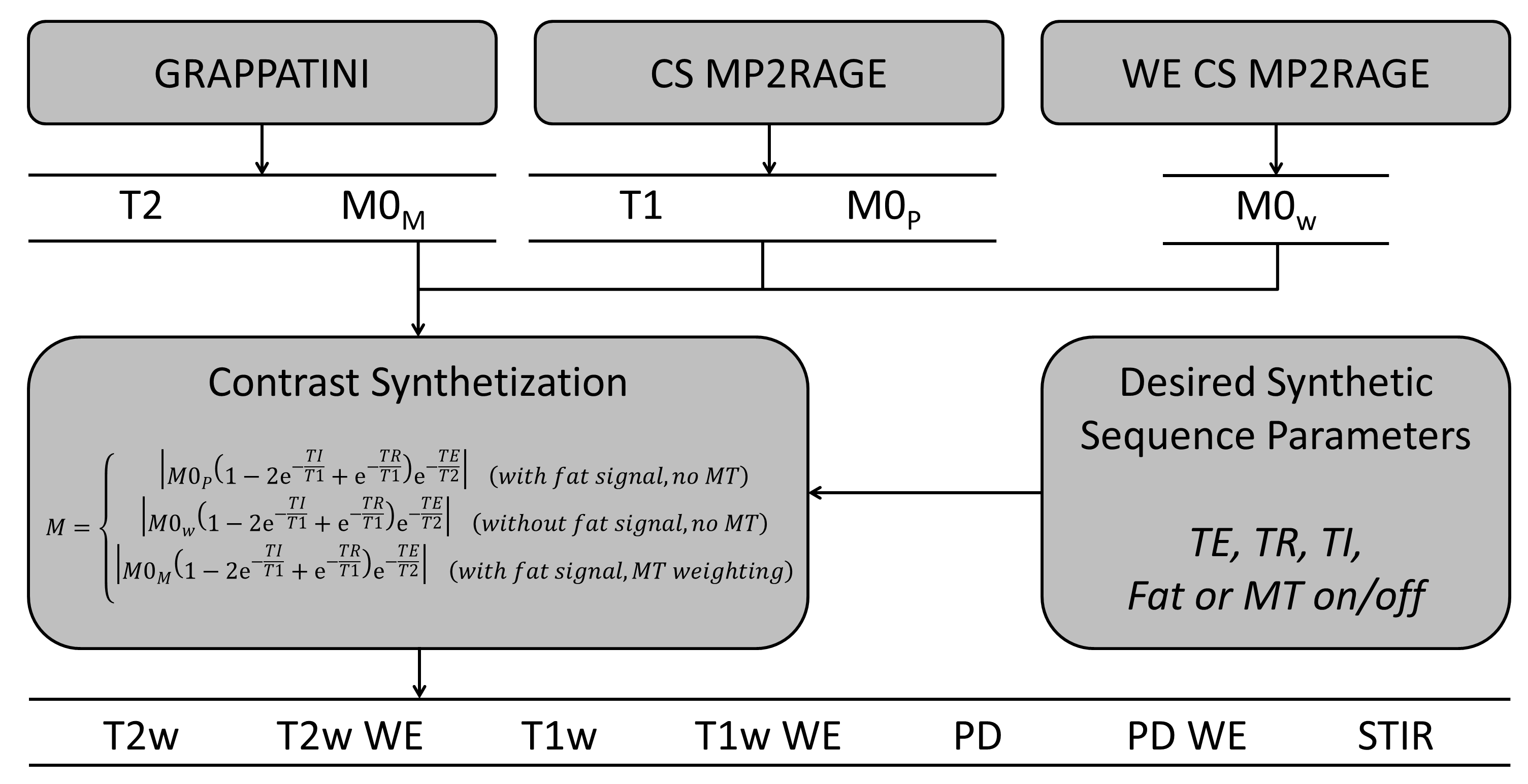
The resulting T1 and T2 maps, and M0P, M0W, M0M images were then used to generate synthetic images M with arbitrary TE, TR, and TI. Additionally, the fat signal or MT-weighting could be turned on and off by switching between M0P, M0W, and M0M:
$$M=\begin{cases}\left|{M0}_P(1-2e^{-\frac{TI}{T1}}+e^{-\frac{TR}{T1}})e^{-\frac{TE}{T2}}\right|&\quad\text{with fat signal, no MT}\\\left|{M0}_W(1-2e^{-\frac{TI}{T1}}+e^{-\frac{TR}{T1}})e^{-\frac{TE}{T2}}\right|&\quad\text{without fat signal, no MT}\\\left|{M0}_M(1-2e^{-\frac{TI}{T1}}+e^{-\frac{TR}{T1}})e^{-\frac{TE}{T2}}\right|&\quad\text{with fat signal,MT weighting}\end{cases}$$
Synthetic contrasts with the same sequence parameters as the conventional turbo spin echo (TSE) pulse sequences were generated and compared visually. Additionally, a series of images was generated by either changing the TE, TR, or TI for fat, non-fat and MT contrasts. Figure 1 shows a schematic flowchart from acquisition to synthetic images.
Results
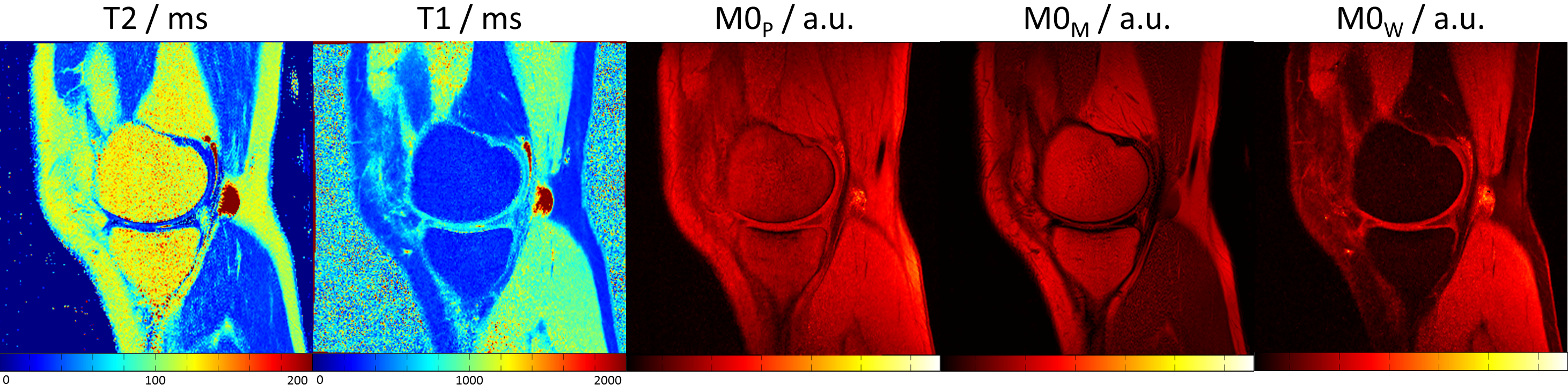
Quantitative maps and the different initial magnetizations from one patient are shown in Figure 2. The M0W shows no fat signal, which, on the contrary, is visible in M0P. When comparing M0M to M0P, it can be seen that especially the muscles show lower signal intensities due to the MT weighting.
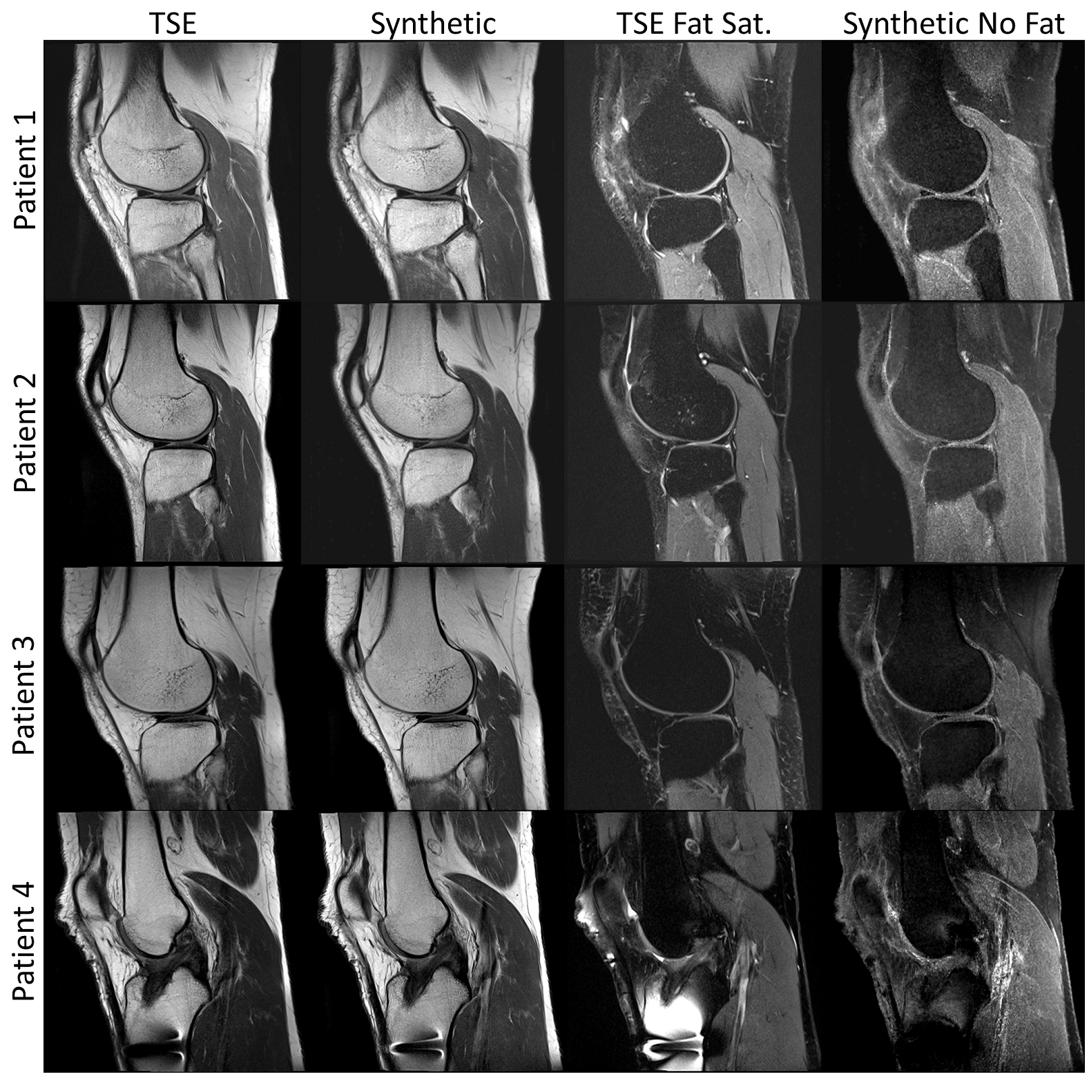
Figure 3 compares the synthetic MR images (with MT weighting and without fat signal) to conventional TSE acquisitions. Visually, the contrasts appear similar, especially for the images with fat signal and MT-weighting. The contrasts without fat signal are similar, but the conventional MR images show higher fluid intensities. The synthetic contrasts without fat signal appear to have more noise in comparison to TSE images.
In Figure 4, multiple parameters are dynamically varied to generate images, showing the entire spectrum of contrasts that can be generated with the proposed method.
Discussion & Conclusion
Our proposed method allows the generation of contrasts with and without fat signal based on quantitative maps. This additional ability of synthetic contrast generation without fat signal may help to limit the introduction of noise that is related to inversion recovery-based techniques, and to diagnose edema lesions, tears, and foci of active inflammation of bone, tendons, ligaments, menisci, muscles, and convective tissues.
Our work was intended as a proof-of-concept and to demonstrate clinical feasibility at 1.5T field strength; however, the signal-to-noise ratio in the synthetic images without fat signal may be improved through protocol optimizations and the use of higher field strengths.
Notably, the synthetic MT-weighting originates from interleaved-slice sampling in the GRAPPATINI sequence and cannot be compared to MT-weighting produced with off-resonance pulses.
For clinical validation, our future work will focus on testing the interchangeability and objectively comparing the image quality of synthetic and conventional MR images. The quantitative maps were previously validated in phantom studies1,3.
In conclusion, we present a fast qMRI method that allows for the synthetic generation of quantitative and qualitative MR images with a wide variety of contrasts, including “on/off” functions for fat and magnetization-transfer-weighting in an acquisition time of 6:46 min for a broad spectrum of MSK applications.
Acknowledgements
No acknowledgement found.References
- Roux M, Hilbert T, Ledoux J-B, Kober T, Omoumi P. Buy one, get two for free: simultaneous knee T2 mapping and morphological analysis on synthetic images using grappatini. Osteoarthr Cartil. 2016;24:S301--S302.
- Kumar NM, Fritz B, Stern SE, et al. Synthetic MRI of the Knee: Phantom Validation and Comparison with Conventional MRI. Radiology. 2018. doi:10.1148/radiol.2018173007.
- Marques JP, Kober T, Krueger G, van der Zwaag W, Van de Moortele PF, Gruetter R. MP2RAGE, a self bias-field corrected sequence for improved segmentation and T1-mapping at high field. Neuroimage. 2010;49(2):1271-1281. doi:10.1016/j.neuroimage.2009.10.002.
- Mussard E, Hilbert T, Meuli R, Thiran J-P, Kober T. Accelerated MP2RAGE imaging using sparse iterative reconstruction. In: ISMRM 2016, ISMRM 24rd Annual Meeting & Exhibition, SMRT 25th Annual Meeting,. ; 2016.
- Hilbert T, Sumpf TJ, Weiland E, et al. Accelerated T 2 mapping combining parallel MRI and model-based reconstruction: GRAPPATINI. J Magn Reson Imaging. 2018. doi:10.1002/jmri.25972.
- Santyr GE. Magnetization transfer effects in multislice MR imaging. Magn Reson Imaging. 1993;11(4):521-532. doi:10.1016/0730-725X(93)90471-O.
Figures